. 2018 Oct 26;9(1):4489.
doi: 10.1038/s41467-018-06794-x.

Common helical V1V2 conformations of HIV-1 Envelope expose the α4β7 binding site on intact virions

Affiliations

Common helical V1V2 conformations of HIV-1 Envelope expose the α4β7 binding site on intact virions

Constantinos Kurt Wibmer et al. Nat Commun. .

Abstract

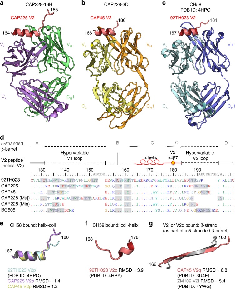
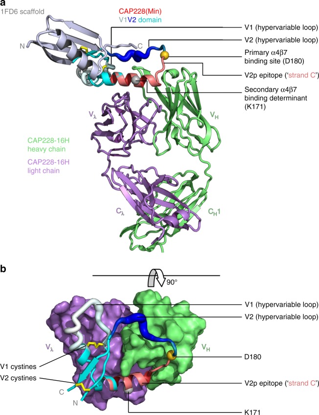
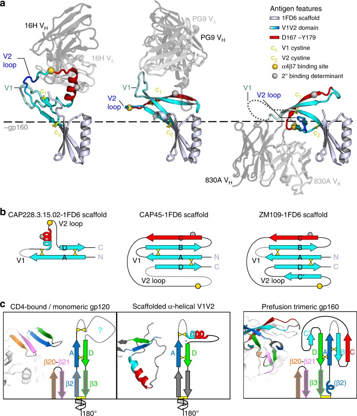
The α4β7 integrin is a non-essential HIV-1 adhesion receptor, bound by the gp120 V1V2 domain, facilitating rapid viral dissemination into gut-associated lymphoid tissues. Antibodies blocking this interaction early in infection can improve disease outcome, and V1V2-targeted antibodies were correlated with moderate efficacy reported from the RV144 HIV-1 vaccine trial. Monoclonal α4β7-blocking antibodies recognise two slightly different helical V2 conformations, and current structural data suggests their binding sites are occluded in prefusion envelope trimers. Here, we report cocrystal structures of two α4β7-blocking antibodies from an infected donor complexed with scaffolded V1V2 or V2 peptides. Both antibodies recognised the same helix-coil V2 conformation as RV144 antibody CH58, identifying a frequently sampled alternative conformation of full-length V1V2. In the context of Envelope, this α-helical form of V1V2 displays highly exposed α4β7-binding sites, potentially providing a functional role for non-native Envelope on virion or infected cell surfaces in HIV-1 dissemination, pathogenesis, and vaccine design.


Conflict of interest statement

The authors declare no competing interests.

Figures

Fig. 1
Fig. 1
HIV-1 V2 peptides reproducibly sample the same helix-coil conformation. a A cartoon representation of the Fab region from V2p mAb CAP228-16H bound to a heterologous V1V2 domain from HIV-1 strain CAP225. Only V2 residues 164–185 were resolved, and are shown in red. The Fab heavy and light chains are coloured green and purple, respectively. b A cartoon representation of the Fab region from V2p mAb CAP228-3D bound to a short, heterologous V2 peptide (shown in red) from HIV-1 strain CAP45. The Fab heavy and light chains are coloured orange and yellow, respectively. c Previously determined cocrystal structure of CH58 bound to the RV144 vaccine strain V2 peptide. d A sequence alignment of the V1V2 domains from the HIV-1 strains for which crystal structures were determined in this study, as well as the dominant (Maj) and subdominant (Min) CAP228 cofounder viruses. The hypervariable loop regions of V1 and V2 are indicated with the dashed lines, while the conserved V2 helix-coil motif is labelled with a red cartoon representation. A gold sphere was used to mark the location of the primary α4β7 binding site, and any potential N-linked glycosylation sites are shaded grey. Above, arrows designate those regions of V1V2 that form the 5-stranded β-barrel bound by V2q/V2i mAbs. e Main chain superimposition of the V2 peptides (residues 167–180) bound by CAP228-16H (purple), CAP228-3D (yellow) and CH58 (light blue). The Cα root-mean-square deviation for each comparison with CH58 bound peptide is shown. f Previously determined structure of a CH59 bound V2 peptide (residues 168–178). g Previously determined structures of the V2 C-sheet in the context of the 5-stranded β-barrel (residues 166–180) when bound by PG9 (red) or 830 A (silver)
Fig. 2
Fig. 2
IGHV5-51 V2p mAbs define a binding class with a distinct mode of recognition. a Logogram of global V2 sequences in the HIV-1 LANL database, showing the relative amino acid frequencies (y-axis) at each position in V2 (x-axis, residues 165-182). Key hydrophobic amino acids bound by IGHV5-51 antibodies are highlighted in darker red, while other key contact residues are coloured black. b A cartoon representation of the three IGHV5-51 V2p Fab paratopes (coloured as in Fig. 1), with the central hydrophobic binding depression shown in surface view. Heavy and light chain residues that make up the hydrophobic binding area are indicated, and the side chains are shown for key buried V2 residues 172, 175 and 176. c The solvent accessible surfaces of the heavy and light chain variable domains from all three mAbs are shown and labelled. The light chains are coloured dark grey, while the heavy chains are coloured on a smoothed charge gradient with more electronegative regions in red, and more electropositive surfaces in blue. The heavy chain complementarity determining regions 1, 2 and 3 are encircled with green, yellow and pink dotted lines, respectively, and the CDR-H3 lengths and overall charges are indicated. V2 peptides are shown in stick and cartoon views. A single rotamer change to account for crystal packing is indicated by the asterisk. d Atomic level details of the interaction between all three IGHV5-51 mAb heavy chains and V2, with the CDRs coloured as in c. Hydrogen bonds are indicated with the dotted black lines, and key water molecules are shown with blue spheres. Alternative rotamer conformations for V1V2 residue D180 and CDR-H1 residue R28 (influenced by crystal packing in the CAP225 bound structure) are shown and labelled with asterisks
Fig. 3
Fig. 3
CAP228 antibodies efficiently inhibit α4β7 integrin binding. a Inhibition of ConC gp120 binding to α4β7 integrin transfected 293 T cells by V2p mAbs CAP228-16H (green), CAP228-3D (orange) or CH58 (blue). Percentage blocking (y-axis) was calculated from the difference in mean fluorescence intensity (MFI) when cell-gp120 complexes were made in the presence or absence of antibody. The α4β7 specific antibodies HP21 (purple) and ACT1 (red) were used as positive controls, while an irrelevant RSV antibody (palivizumab) and an HIV-1 CD4 binding site-specific antibody (VRC01) were used as the negative controls. b Ability of V2p antibodies CAP228-16H (green) or CH58 (blue) to block adhesion of α4β7 integrin expressing RPMI8866 cells to a cyclic 92TH023 peptide (residues 157–196) at three separate concentrations. c Inhibition of V2 peptide (strains 92TH023 or BG505) binding to α4β7 integrin expressing RPMI8866 cells by V2p mAbs CAP228-16H (green), CAP228-3D (orange), CH58 (blue) and CH59 (light blue) or the conformation specific non-V2p mAb CAP228-9D. The mAb 2B4 is specific for the α4 integrin and served as the positive control (pink). Data are represented as the result of up to three biological replicates used to calculate a mean
Fig. 4
Fig. 4
Cocrystal structure of a scaffolded, helical V1V2 domain bound to CAP228-16H. a The 1FD6 scaffold (metallic blue), autologous CAP228(Min) V1V2 domain (multicolour) and CAP228-16H Fab heavy and light chains (green and purple, respectively) are shown in cartoon view. V1V2 regions are labelled as hypervariable loop 1 (residues 132–156, white), hypervariable loop 2 (residues 183–196, dark blue), and C strand (residues 167–179, red), with the remainder coloured cyan. Internal V1V2 disulphide bonds are shown as yellow sticks, while the primary α4β7 binding site and potential secondary α4β7 binding determinant are indicated with the gold and silver spheres, respectively. b The view has been rotated 90°, and the 1FD6 scaffold (which would point ‘out of the page’) has been omitted to clearly show the interaction. The CAP228-16H paratope is shown in surface view, with the V1V2 domain still in cartoon representation, coloured as above
Fig. 5
Fig. 5
Comparisons of the V1V2 domain when bound by V2p, V2q and V2i mAbs. a A cartoon representation of CAP228-16H (V2p, left), PG9 (V2q, middle) and 830 A (V2i, right) bound to 1FD6 scaffolded V1V2. Only the Fab variable domains are shown for clarity, with the heavy and light chains coloured dark and light washed-out grey, respectively. All three structures are oriented with respect to the 1FD6 scaffold (metallic blue). Residues 167–179 that make up the V1V2 C-strand, or the IGHV5-51 bound α-helix, are shown in bright red, with the remaining V1V2 regions coloured as in Fig. 4. The V1V2 internal disulphide bonds are shown with yellow sticks, and α4β7 binding determinants are labelled with gold and silver spheres. A dashed line was used to indicate the approximate location for the rest of gp160 relative to full length V1V2. b Schematic of the V1V2 domain, showing the relative refolding of B- and C-strands, and repositioning of the A- and D-strands between the α-helical and β-stranded conformations of V1V2. The α4β7 binding determinants and disulphide bonds are labelled as in a. c Cartoon views and schematic representations of V1V2 in the CD4-bound or monomeric gp160 state (left panel), the V2p-bound α-helical state (middle panel), and the prefusion, entry-competent gp160 trimer (right panel). The β20 and β21 strands that make up one half of the bridging sheet are coloured orange and pink, respectively. The β2 strand of gp120 (or overlapping prefusion associated α-helical structure), and subsequent V1V2 A-strand region is coloured dark blue, while the V1V2 D-strand and subsequent β3 strand of gp120 is coloured green. The remaining V1V2 region is coloured as in b
Fig. 6
Fig. 6
The α-helical form of V1V2 potentially engages the α4β7 integrin with high efficacy. a Cell surface staining of HIV-1 infected CD4+ T cells (strains 92TH023 and CMU06) using RV144 mAbs CH58 (left) and CH59 (right). An uninfected control (top panels) was included to estimate background and showed minimal (<1%) staining of cells. Plots are shown as forward scatter (cell size indicator) vs. level of phycoerythrin conjugated staining of either mAb. b Endpoint ELISA binding titres of V2p mAbs CAP228-16H (green), CAP228-3D (orange) or CH59 (light blue), quaternary structure dependent V2q mAb PGT145 (purple) or V2i mAb 830 A (yellow) to concentrated pseudovirus expressing the C1080 envelope. HIV-1 CD4 binding site-specific antibody VRC01, and an irrelevant Flu-specific antibody were used as positive or negative controls, respectively. c Models of the HIV-1 prefusion Env trimer (left), misfolded/refolded helical V1V2 (centre) or gp160 that has adopted the CD4-bound conformation (right) are shown. The latter two conformations of Env display highly exposed α4β7 binding sites (gold spheres) within helical V1V2, providing a potentially functional role for aberrant Env on the viral surface in α4β7 integrin binding and viral dissemination

Similar articles

Cited by

References

    1. Leonard CK, et al. Assignment of intrachain disulfide bonds and characterization of potential glycosylation sites of the type 1 recombinant human immunodeficiency virus envelope glycoprotein (gp120) expressed in Chinese hamster ovary cells. J. Biol. Chem. 1990;265:10373–10382. - PubMed
    1. Mao Youdong, Wang Liping, Gu Christopher, Herschhorn Alon, Xiang Shi-Hua, Haim Hillel, Yang Xinzhen, Sodroski Joseph. Subunit organization of the membrane-bound HIV-1 envelope glycoprotein trimer. Nature Structural & Molecular Biology. 2012;19(9):893–899. doi: 10.1038/nsmb.2351. - DOI - PMC - PubMed
    1. Pancera M, et al. Structure and immune recognition of trimeric pre-fusion HIV-1 Env. Nature. 2014;514:455–461. doi: 10.1038/nature13808. - DOI - PMC - PubMed
    1. Julien JP, et al. Crystal structure of a soluble cleaved HIV-1 envelope trimer. Science. 2013;342:1477–1483. doi: 10.1126/science.1245625. - DOI - PMC - PubMed
    1. Bartesaghi A, Merk A, Borgnia MJ, Milne JL, Subramaniam S. Prefusion structure of trimeric HIV-1 envelope glycoprotein determined by cryo-electron microscopy. Nat. Struct. Mol. Biol. 2013;20:1352–1357. doi: 10.1038/nsmb.2711. - DOI - PMC - PubMed

Publication types

MeSH terms