

Protein-Engineered Large Area Adipose-derived Stem Cell Sheets for Wound Healing
- PMID: 30367098
- PMCID: PMC6203842
- DOI: 10.1038/s41598-018-34119-x
Protein-Engineered Large Area Adipose-derived Stem Cell Sheets for Wound Healing
Abstract
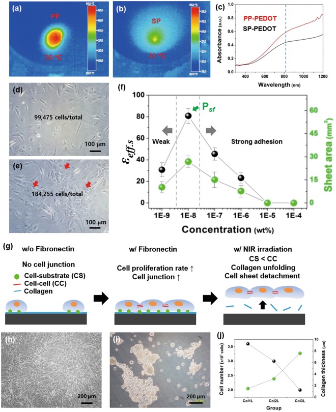
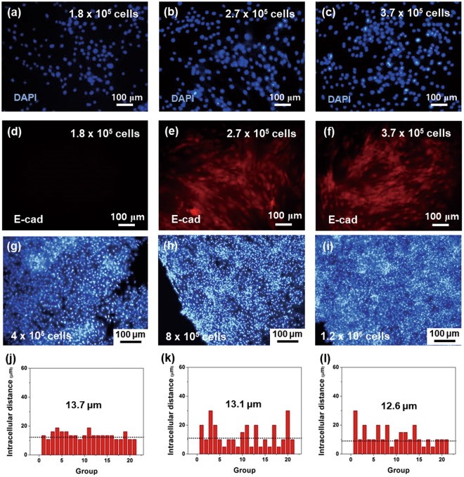
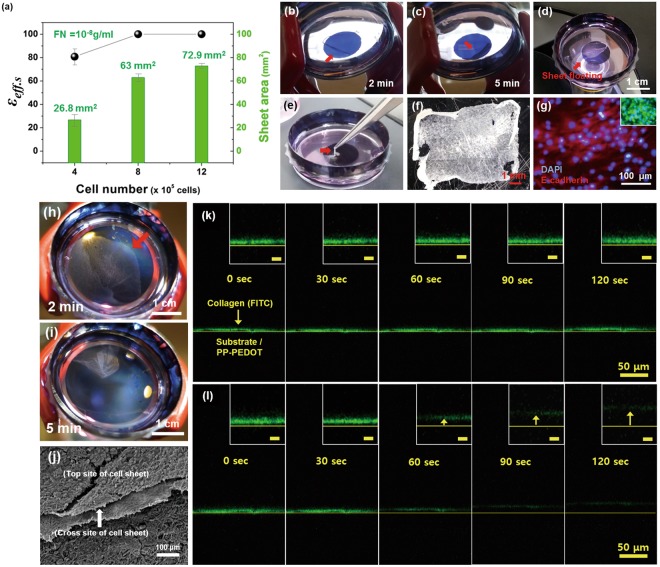
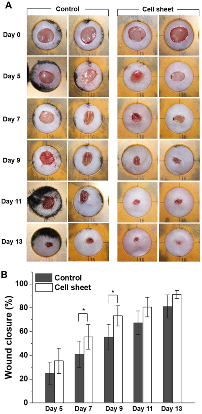
Human adipose-derived stem cells (hADSCs) formed robust cell sheets by engineering the cells with soluble cell adhesive molecules (CAMs), which enabled unique approaches to harvest large area hADSC sheets. As a soluble CAM, fibronectin (FN) (100 pg/ml) enhanced the cell proliferation rate and control both cell-to-cell and cell-to-substrate interactions. Through this engineering of FN, a transferrable hADSC sheet was obtained as a free-stranding sheet (122.6 mm2) by a photothermal method. During the harvesting of hADSC sheets by the photothermal method, a collagen layer in-between cells and conductive polymer film (CP) was dissociated, to protect cells from direct exposure to a near infrared (NIR) source. The hADSC sheets were applied to chronic wound of genetically diabetic db/db mice in vivo, to accelerate 30% faster wound closure with a high closure effect (εwc) than that of control groups. These results indicated that the engineering of CAM and collagens allow hADSC sheet harvesting, which could be extended to engineer various stem cell sheets for efficient therapies.
Conflict of interest statement
The authors declare no competing interests.
Figures
Similar articles
-
Effective stacking and transplantation of stem cell sheets using exogenous ROS-producing film for accelerated wound healing.Acta Biomater. 2019 Sep 1;95:418-426. doi: 10.1016/j.actbio.2019.01.019. Epub 2019 Jan 16. Acta Biomater. 2019. PMID: 30660002
-
Engineering, differentiation and harvesting of human adipose-derived stem cell multilayer cell sheets.Regen Med. 2019 Mar;14(3):151-163. doi: 10.2217/rme-2018-0053. Epub 2019 Mar 4. Regen Med. 2019. PMID: 30829557
-
Creation and Transplantation of an Adipose-derived Stem Cell (ASC) Sheet in a Diabetic Wound-healing Model.J Vis Exp. 2017 Aug 4;(126):54539. doi: 10.3791/54539. J Vis Exp. 2017. PMID: 28809824 Free PMC article.
-
Mesenchymal Stem Cells Improve Healing of Diabetic Foot Ulcer.J Diabetes Res. 2017;2017:9328347. doi: 10.1155/2017/9328347. Epub 2017 Mar 12. J Diabetes Res. 2017. PMID: 28386568 Free PMC article. Review.
-
Stem cell therapies for wound healing.Expert Opin Biol Ther. 2019 Jun;19(6):575-585. doi: 10.1080/14712598.2019.1596257. Epub 2019 Apr 11. Expert Opin Biol Ther. 2019. PMID: 30900481 Review.
Cited by
-
Effect of composite biodegradable biomaterials on wound healing in diabetes.Front Bioeng Biotechnol. 2022 Nov 25;10:1060026. doi: 10.3389/fbioe.2022.1060026. eCollection 2022. Front Bioeng Biotechnol. 2022. PMID: 36507270 Free PMC article. Review.
-
Autologous adipose-derived stem cell transplantation enhances healing of wound with exposed bone in a rat model.PLoS One. 2019 May 13;14(5):e0214106. doi: 10.1371/journal.pone.0214106. eCollection 2019. PLoS One. 2019. PMID: 31083652 Free PMC article.
-
The preclinical and clinical progress of cell sheet engineering in regenerative medicine.Stem Cell Res Ther. 2023 Apr 27;14(1):112. doi: 10.1186/s13287-023-03340-5. Stem Cell Res Ther. 2023. PMID: 37106373 Free PMC article. Review.
-
Current status and future prospects of stomatology research.J Zhejiang Univ Sci B. 2023 Aug 7;24(10):853-867. doi: 10.1631/jzus.B2200702. J Zhejiang Univ Sci B. 2023. PMID: 37752088 Free PMC article.
-
Fibronectin in hyperglycaemia and its potential use in the treatment of diabetic foot ulcers: A review.Int Wound J. 2023 May;20(5):1750-1761. doi: 10.1111/iwj.13997. Epub 2022 Dec 19. Int Wound J. 2023. PMID: 36537075 Free PMC article. Review.
References
-
- McFarlin Kellie, Gao Xiaohua, Liu Yong Bo, Dulchavsky Deborah S., Kwon David, Arbab Ali S., Bansal Mona, Li Yi, Chopp Michael, Dulchavsky Scott A., Gautam Subhash C. Bone marrow-derived mesenchymal stromal cells accelerate wound healing in the rat. Wound Repair and Regeneration. 2006;14(4):471–478. doi: 10.1111/j.1743-6109.2006.00153.x. - DOI - PubMed
Publication types
MeSH terms
Substances
Grants and funding
- 2016K1A1A2912753/National Research Foundation of Korea (NRF)/International
- 2016K1A1A2912753/National Research Foundation of Korea (NRF)/International
- 2016K1A1A2912753/National Research Foundation of Korea (NRF)/International
- 2016K1A1A2912753/National Research Foundation of Korea (NRF)/International
- HI15C0942/Korea Health Industry Development Institute (KHIDI)/International
- HI15C0942/Korea Health Industry Development Institute (KHIDI)/International
- HI15C0942/Korea Health Industry Development Institute (KHIDI)/International
- HI15C0942/Korea Health Industry Development Institute (KHIDI)/International
- HI15C0942/Korea Health Industry Development Institute (KHIDI)/International
- HI15C0942/Korea Health Industry Development Institute (KHIDI)/International
- HI15C0942/Korea Health Industry Development Institute (KHIDI)/International
- HI15C0942/Korea Health Industry Development Institute (KHIDI)/International
- HI15C0942/Korea Health Industry Development Institute (KHIDI)/International
LinkOut - more resources
Full Text Sources
Molecular Biology Databases
Research Materials
Miscellaneous
