. 2018 Oct 26;8(1):15855.
doi: 10.1038/s41598-018-34193-1.

Using patient-derived iPSCs to develop humanized mouse models for chronic myelomonocytic leukemia and therapeutic drug identification, including liposomal clodronate

Affiliations

Using patient-derived iPSCs to develop humanized mouse models for chronic myelomonocytic leukemia and therapeutic drug identification, including liposomal clodronate

Kazuki Taoka et al. Sci Rep. .

Abstract

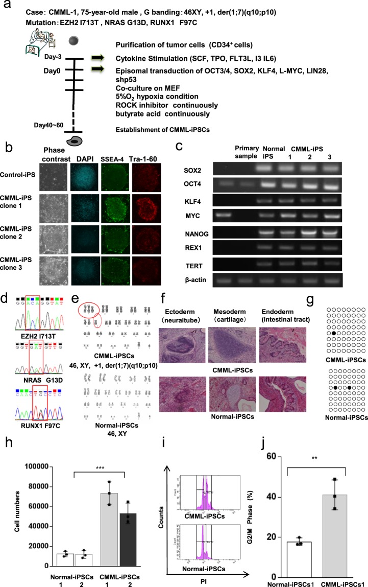
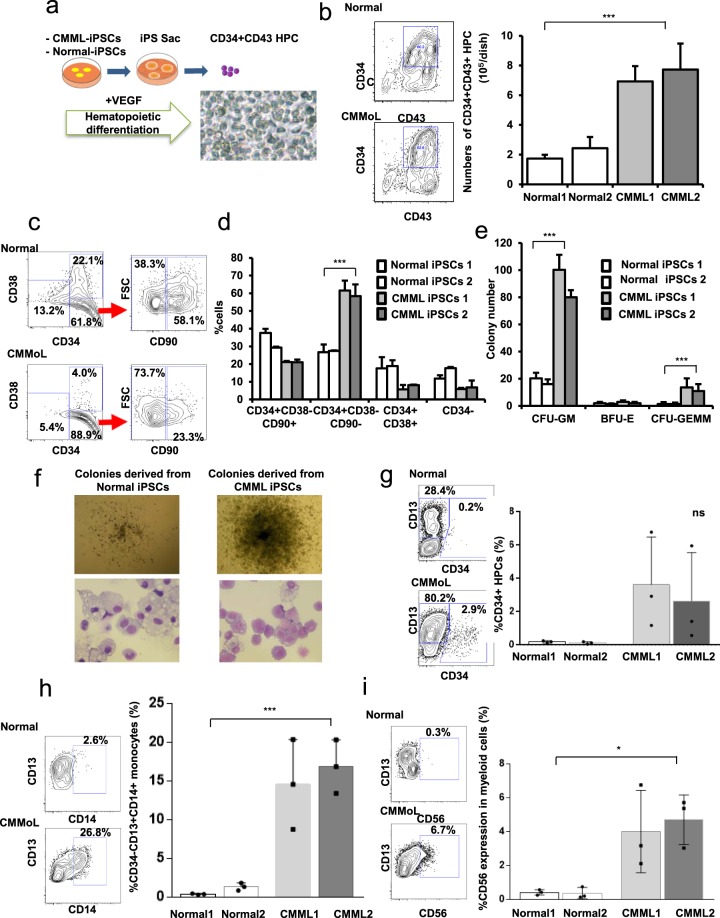
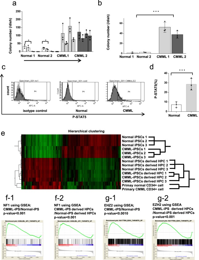
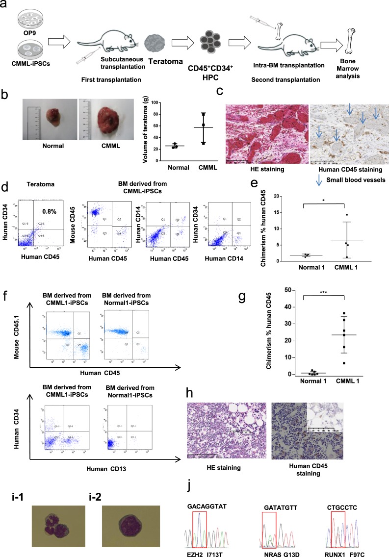
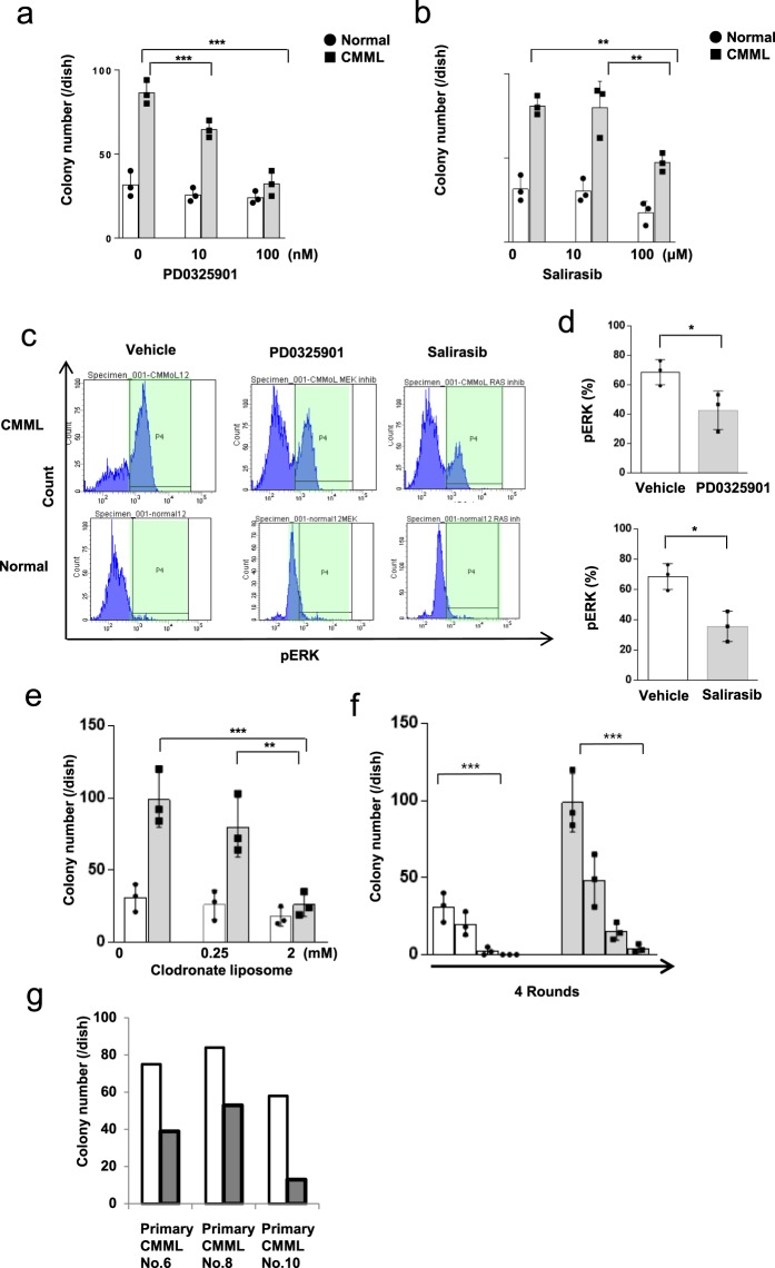
Chronic myelomonocytic leukemia (CMML) is an entity of myelodysplastic syndrome/myeloproliferative neoplasm. Although CMML can be cured with allogeneic stem cell transplantation, its prognosis is generally very poor due to the limited efficacy of chemotherapy and to the patient's age, which is usually not eligible for transplantation. Comprehensive analysis of CMML pathophysiology and the development of therapeutic agents have been limited partly due to the lack of cell lines in CMML and the limited developments of mouse models. After successfully establishing patient's derived disease-specific induced pluripotent stem cells (iPSCs) derived from a patient with CMML, we utilized these CMML-iPSCs to achieve hematopoietic re-differentiation in vitro, created a humanized CMML mouse model via teratomas, and developed a drug-testing system. The clinical characteristics of CMML were recapitulated following hematopoietic re-differentiation in vitro and a humanized CMML mouse model in vivo. The drug-testing system using CMML-iPSCs identified a MEK inhibitor, a Ras inhibitor, and liposomal clodronate as potential drugs for treating CMML. Clodronate is a drug commonly used as a bisphosphonate for osteoporosis. In this study, the liposomalization of clodronate enhanced its effectiveness in these assays, suggesting that this variation of clodronate may be adopted as a repositioned drug for CMML therapy.


Conflict of interest statement

The authors declare no competing interests.

Figures

Figure 1
Figure 1
Generation of CMML patient-derived iPSCs. (a) Protocol for the generation of CMML patient-derived iPSCs. CD34+ cells from patient samples were isolated from BM mononuclear cells. OCT3/4, SOX2, KLF4, L-MYC, LIN 28, and shP53 were transduced using episomal vectors under hypoxic conditions in the presence of a Rho kinase (ROCK) inhibitor and butyrate acid. Three clones of CMML iPSCs from one patient with CMML-1 were established. (b) Immunofluorescence staining of pluripotency marker antigens (SSEA-4 and Tra-1-60) in Normal and CMML iPSCs. (c) Semi-quantitative RT-PCR of pluripotency markers. The endogenous expression of pluripotent stem cell-specific genes (OCT3/4, SOX2, KLF4, C-MYC, NANOG, REX1, and TERT) was confirmed. Each of the images is cropped from different gels. (d) EZH2, RUNX1, and NRAS mutations were identified in CMML iPSCs. (e) Representative karyotypes of CMML iPSCs showing derivative chromosome (1;7)(q10;p10), an unbalanced translocation, and Normal-iPSCs. (f) Histological analyses of teratomas from CMML iPSCs. A teratoma with three germ layers, the ectoderm (neural tube), mesoderm (cartilage), and endoderm (intestinal tract), was observed following H&E staining. (g) Bisulfite sequence analysis of the NANOG gene promoter; the black circles represent methylated CpG, while the white circles represent unmethylated CpG. (h) CMML iPSCs grew rapidly and displayed a five-fold higher proliferation rate compared to control iPSCs (n = 3 independent experiments, ***p < 0.001, CMML-iPSCs 2 clones and control iPSCs 2 clones derived from same donor samples, paired two-sided t-test,). (ij) Cell cycle analysis showed a relative increase in CMML iPSCs in the G2/M phase. Statistical analysis was performed (n = 3 independent experiments, **p < 0.01, CMML-iPSCs 1 clones and control iPSCs 1 clones derived from same donor samples, Student’s t test). Horizontal lines represent the means ± s.d.
Figure 2
Figure 2
The patient’s pathogenesis of CMML was recapitulated in vitro in CMML iPSC-derived hematopoietic progenitor cells (HPCs). (a) Scheme for inducing CMML and Normal iPSC-derived HPCs. We obtained CD34 + CD43 + hematopoietic progenitor cells in CMML-iPS-sac on day 17 of the co-culture system. We evaluated two different lines of one normal and unique one CMML iPSC clone in triplicate tests. (b) CMML iPSCs generated more CD34+ CD43+ HPCs than Normal-iPSCs (n = 3, independent experiments, ***p < 0.001, Normal-iPSCs1 and 2 from a healthy donor, and CMML-iPSCs1 and 2 from a CMML patient, paired two-sided t-test). (c,d) The CD34+ CD38 CD90fraction re-differentiated from CMML-iPSCs increased more than Normal-iPSCs (n = 3, independent experiments, ***p < 0.001, Normal-iPSCs1 and 2 from a healthy donor, and CMML-iPSCs1 and 2 from a CMML patient, Experiments were performed in triplicate. Statistical analyses were performed with ANOVA and the Dunnett post-test for multiple comparisons. ***p < 0.001). (e) The semi-solid culture of CD34+ CD43+ HPCs derived from CMML-iPSCs yielded many more CFU-GM and CFU-GEMM colonies, compared to their normal counterparts (n = 3, independent experiments, ***p < 0.001, Normal-iPSCs1 and 2 from a healthy donor, and CMML-iPSCs1 and 2 from a CMML patient, Statistical analyses were performed with ANOVA and the Dunnett post-test for multiple comparisons. ***p < 0.001). (f) Colonies derived from CMML iPSCs were larger compared to those from Normal-iPSCs. (gi) The profiles of the surface antigens in the colonies from CMML iPSC-derived HPCs and controls were analyzed using FACS (n = 3, independent experiments, *p < 0.05,***p < 0.001, Normal-iPSCs1 and 2 from a healthy donor, and CMML-iPSCs1 and 2 from a CMML patient, paired two-sided t-test). Horizontal lines represent the means ± s.d.
Figure 3
Figure 3
Characteristics of colony formation and genetic analysis of CMML iPSC-derived HPCs. (a) CMML iPSC-derived HPCs retained their serial re-plating capacity and generated colonies even after the fourth plating, whereas Normal iPSC-derived HPCs lost the ability to generate colonies after the second plating. CMML iPSC-derived HPCs showed an enhanced self-renewal capacity (n = 3, independent experiments, *p < 0.05, Normal-iPSCs1 and 2 from a healthy donor, and CMML-iPSCs1 and 2 from a CMML patient, Statistical analyses were performed with ANOVA and the Dunnett post-test for multiple comparisons. *p < 0.05). (b) The CMML iPSC-derived HPCs generated hematopoietic colonies under cytokine-deprived conditions. The CMML iPSC-derived HPCs could form hematopoietic colonies, while Normal iPSC-derived cells gave rise to few detectable colonies (n = 3, independent experiments, ***p < 0.001, Normal-iPSCs1 and 2 from a healthy donor, and CMML-iPSCs1 and 2 from a CMML patient, paired two-sided t-test). (c) Using pStat5 intracellular flow cytometric analysis, we observed increased phosphorylation of STAT5 in CMML iPSC-derived CD34+ CD43+ HPCs. (gi) The profiles of the surface antigens in the colonies from CMML iPSC-derived HPCs and controls were analyzed using FACS. (d) The percentage of pSTAT5 on CMML iPSC-derived CD34+ CD43+ HPCs was significantly higher than Normal- iPSC-derived HPCs (n = 3, independent experiments, ***p < 0.001, Normal-iPSCs1 from a healthy donor, and CMML-iPSCs1 from a CMML patient, paired two-sided t-test). (e) Hierarchical clustering analysis of gene expression patterns. Using hierarchical clustering, these cells were distinguished into two groups: the Normal/CMML iPSC group and Normal/CMML iPSC-derived HPC group. (f-1 and f-2) Expression data were analyzed to identify genes in CMML-iPSC-derived HPCs and CMML-iPSCs that positively affected NF1 using GSEA.(p < 0.001). (g-1 and g-2) Expression data were analyzed to identify genes in CMML-iPSC-derived HPCs and CMML-iPSCs that positively affect EZH2 using GSEA. (p < 0.001). Horizontal lines represent the means ± s.d.
Figure 4
Figure 4
In vivo CMML xenograft model recapitulate its pathogenesis. (a) Experimental scheme for the first transplantation. We co-injected CMML-iPSCs along with OP9 stromal cells into immunodeficient mice using an in vivo system in which human CMML iPSCs develop into HPCs within teratomas. Second transplantation of CMML progenitor cells generated in vivo. (b) CMML iPS-induced teratomas had larger volumes than the induced teratomas from Normal-iPSCs (n = 3, independent experiments, Normal-iPSCs1 from a healthy donor, and CMML-iPSCs 1 from a CMML patient, paired two-sided t-test). (c) Immunostaining of CD45+ blood cells around the small blood vessels generated in the teratoma. (d) CD45+ CD34+ HPCs were isolated from the teratoma. Leakage of the CD45+ CMML cells from the teratoma into the BM was observed. (e) Estimated number of CMML cells in the BM of engrafted mice in which teratomas formed from tCMML iPS cells (n = 4, independent experiments, *p < 0.05, Normal-iPSCs1 from a healthy donor, and CMML-iPSC1 from a CMML patient, paired two-sided t-test). (f) CMML-iPSC-derived blood in vivo reproduced and recapitulated the profiles of CMML surface antigens, CD13+ or CD34+. (g) Flow cytometry revealed human chimerism of CD45+ cells in the BM of the secondarily transplanted NSG mice. Statistical analysis was performed (n = 6, independent experiments, *p < 0.05, Normal-iPSCs1 from a healthy donor, and CMML-iPSCs1 from a CMML patient, paired two-sided t-test). (h) H&E staining and anti-human CD45 immunostaining. (i) Morphological analysis revealed that the sorted CD45+ human cells consisted of monocytes and monoblasts (i-1) and a few blasts (i-2). (j) EZH2, RUNX1, and NRAS mutations identified in the BM of secondarily transplanted NSG mice. Horizontal lines represent the means ± s.d.
Figure 5
Figure 5
Drug discovery using CMML-iPSCs. (a) An MEK inhibitor (PD0325901) reduced the colony-forming capacity of CMML iPSC-derived HPCs (n = 3, independent experiments,, Normal-iPSCs1 from a healthy donor, and CMML-iPSCs1 from a CMML patients, Experiments were performed in triplicate. Statistical analyses were performed with ANOVA and the Dunnett post-test for multiple comparisons. ***p < 0.001)). (b) RAS inhibitor (salirasib) reduced the colony-forming capacity of CMML iPSC-derived HPCs (n = 3, independent experiments, Normal-iPSCs1 from a healthy donor, and CMML-iPSCs1 from a CMML patients, Experiments were performed in triplicate. Statistical analyses were performed with ANOVA and the Dunnett post-test for multiple comparisons. **p < 0.01). (c) pERK was constitutively activated only in the colonies of CMML iPSC-derived HPCs. Furthermore, ERK phosphorylation was inhibited by MEK and RAS inhibitors in these colonies. (d) The expression level of pERK was significantly decreased in CMML-iPSC-derived hematopoietic colonies by MEK or Ras inhibitor (n = 3, independent experiments, *p < 0.05, Normal-iPSCs1 from a healthy donor, and CMML-iPSCs1 from a CMML patients, paired two-sided t-test). (e) Liposomal clodronate suppressed the growth of colonies from CMML iPSCs. Statistical analysis was performed (n = 3, independent experiments, Normal-iPSCs1 from a healthy donor, and CMML-iPSCs1 from a CMML patients, Experiments were performed in triplicate. Statistical analyses were performed with ANOVA and the Dunnett post-test for multiple comparisons. ***p < 0.001, **p < 0.01,). (f) Liposomal clodronate reduced re-plating colonies formed by CMML iPSC-derived HPCs (n = 3, independent experiments, Normal-iPSCs1 from a healthy donor, and CMML-iPSCs1 from a CMML patients, Experiments were performed in triplicate. Statistical analyses were performed with ANOVA and the Dunnett post-test for multiple comparisons. ***p < 0.001). (g) Colony formation by three other primary samples from CMML patients was reduced by liposomal clodronate. Horizontal lines represent the means ± s.d.

Similar articles

Cited by

References

    1. Takahashi K, et al. Induction of pluripotent stem cells from adult human fibroblasts by defined factors. Cell. 2007;131:861–872. doi: 10.1016/j.cell.2007.11.019. - DOI - PubMed
    1. Yilmazer A, de Lazaro I, Taheri H. Reprogramming cancer cells: a novel approach for cancer therapy or a tool for disease-modeling? Cancer Lett. 2015;369:1–8. doi: 10.1016/j.canlet.2015.06.027. - DOI - PubMed
    1. Arai S, Miyauchi M, Kurokawa M. Modeling of hematologic malignancies by iPS technology. Exp Hematol. 2015;43:654–660. doi: 10.1016/j.exphem.2015.06.006. - DOI - PubMed
    1. Kotini AG, et al. Functional analysis of a chromosomal deletion associated with myelodysplastic syndromes using isogenic human induced pluripotent stem cells. Nat Biotechnol. 2015;33:646–655. doi: 10.1038/nbt.3178. - DOI - PMC - PubMed
    1. Kotini AG, et al. Stage-Specific Human Induced Pluripotent Stem Cells Map the Progression of Myeloid Transformation to Transplantable Leukemia. Cell Stem Cell. 2017;20:315–328.e317. doi: 10.1016/j.stem.2017.01.009. - DOI - PMC - PubMed

MeSH terms