. 2019 Jan;257(1):83-94.
doi: 10.1007/s00417-018-4166-7. Epub 2018 Oct 26.

Fate of the Fc fusion protein aflibercept in retinal endothelial cells: competition of recycling and degradation

Affiliations

Fate of the Fc fusion protein aflibercept in retinal endothelial cells: competition of recycling and degradation

Heidrun L Deissler et al. Graefes Arch Clin Exp Ophthalmol. 2019 Jan.

Abstract

Purpose: Intravitreal injection of the VEGF-binding protein aflibercept is widely used to treat various ocular diseases. In vitro, immortalized bovine retinal endothelial cells (iBREC) take up and transport aflibercept through the cell layer in a serum-dependent manner, likely mediated through the neonatal Fc receptor (FcRn), but degradation of the Fc domain-containing protein might be a competing intracellular process. Therefore, aflibercept's associations with proteins either involved in FcRn-mediated transport or in the lysosomal pathway were studied.

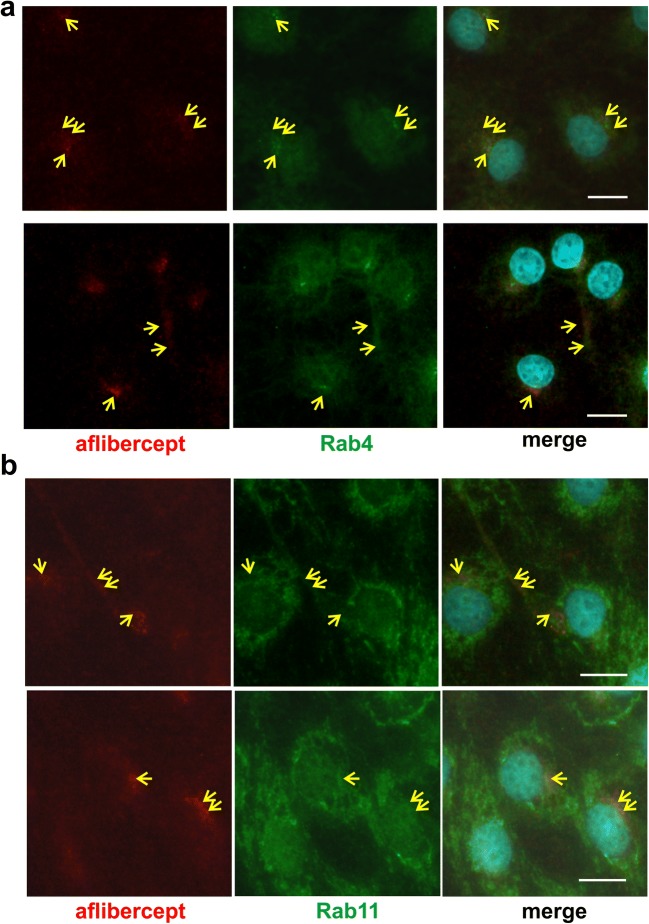
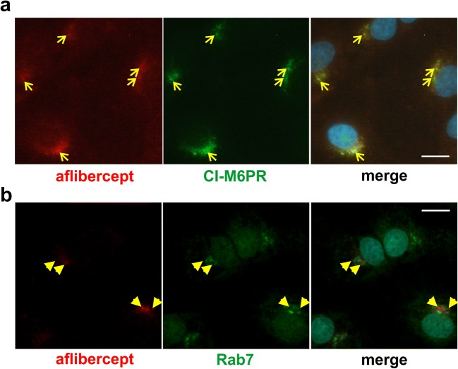
Methods: Confluent iBREC pre-cultivated with or without FBS were exposed for 4 h to in vivo achievable 250 μg/ml aflibercept, before cells were harvested for immunofluorescence staining or preparation of protein extracts. Intracellular localization of aflibercept and putative co-localizations with proteins involved in transport of IgG/FcRn complexes, i.e., endosomal Rab4 and Rab11, components of the cytoskeleton, motor proteins, or with marker proteins characteristic of multivesicular bodies or lysosomes were assessed by co-immunofluorescence stainings. Amounts of expressed endogenous proteins and of internalized aflibercept were determined by Western blot analyses.

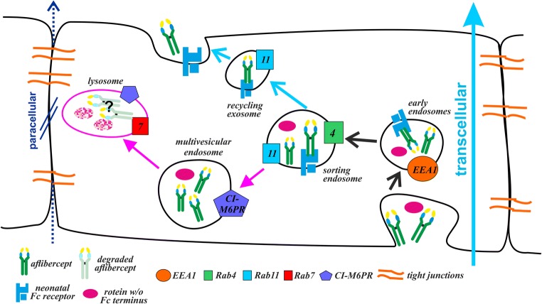
Results: Aflibercept-specific perinuclear staining overlapped with that of the motor protein dynein whereas double staining with an anti-kinesin antibody resulted in a patchy pattern. In addition, aflibercept was typically present close to microtubules and often co-localized with α-tubulin. Rab4 and Rab11 stainings partly overlapped with the perinuclear staining of aflibercept whereas co-localization with Rab7 (in late endosomes/lysosomes) was only rarely seen. Interestingly, aflibercept but not the IgG bevacizumab broadly co-localized with the cation-independent mannose 6-phosphate receptor characteristic of multivesicular endosomes. In accordance with partial degradation beside transcytosis, the amount of intracellular aflibercept increased when cells were treated with protease inhibitors MG-132 or MG-101. Serum-deprived iBREC expressed less Rab11 and dynein but slightly more Rab4.

Conclusion: After uptake by iBREC, aflibercept is present in organelles associated with FcRn-mediated transport, but part of the protein is subject to degradation. Transport inhibition of aflibercept during cultivation without FBS is likely a consequence of an attenuated exocytosis due to decreased expression of Rab11.

Keywords: Aflibercept; Degradation; Fc fusion protein; IgG; Neonatal Fc receptor; Recycling; Retinal endothelial cells.


Conflict of interest statement

Conflict of interest

Heidrun L. Deissler has received funding from Novartis Pharma GmbH, Bayer Vital GmbH, and lecture fees from Bayer Vital GmbH. Gerhard K. Lang has received honoraria from Bayer Vital GmbH. Gabriele E. Lang has received funding from Novartis Pharma GmbH, Bayer Vital GmbH, Boehringer Ingelheim Pharma, Allergan, and Alcon Pharma GmbH and has received honoraria as a consultant from Boehringer Ingelheim Pharma and as a consultant and speaker from Novartis Pharma GmbH, Carl Zeiss Meditec, Alimera Sciences, and Bayer Vital GmbH.

Ethical approval

This article does not contain any studies with human participants or animals performed by any of the authors.

Figures

Fig. 1
Fig. 1
Aflibercept is co-localized with α-tubulin and dynein. Confluent iBREC treated with aflibercept were immunostained to detect aflibercept (red), α-tubulin (a, green), dynein intermediate chain IC74 (b, green), or kinesin heavy chain (HC, c, green). a α-Tubulin fibers originating from the microtubule organization center stretched to the periphery of the cells. Aflibercept-specific signals overlapped with those of α-tubulin at the microtubule organization center (yellow arrows). They were also close to the α-tubulin fibers in the cell periphery (blue arrows). b Dynein signals around the nucleus overlapped with those of aflibercept (yellow arrows). c The strong perinuclear kinesin-specific staining and that of aflibercept gave rise to a mosaic pattern (yellow arrowheads). Scale bar, 10 μm
Fig. 2
Fig. 2
Aflibercept is co-localized with Rab4 and Rab11. Aflibercept (red), Rab4 (a, green), or Rab11 (b, green) were visualized by double immunofluorescence staining after exposure of iBREC to the Fc fusion protein. Antibodies specific for Rab4 (a) or Rab11 (b) bound to a prominent perinuclear region stretching into the periphery of the cell with similar but not identical patterns. Overlapping of signals (yellow arrows) from Rab4 (a) or Rab11 (b) staining with those specific for aflibercept is most evident in the perinuclear region and partially seen in the peripheral areas. Scale bar, 10 μm
Fig. 3
Fig. 3
Aflibercept is largely co-localized with CI-M6PR. iBREC exposed to aflibercept were immunostained to detect aflibercept (red) and either the marker of multivesicular endosomes CI-M6PR (a, green) or Rab7 (b, green), specific for late endosomes and lysosomes. a The prominent CI-M6PR-specific staining close to the nucleus broadly overlapped with that of aflibercept (yellow arrows). b In contrast, aflibercept-specific signals and those specific for Rab7 gave rise to a mosaic pattern with few overlaps (yellow arrowheads). Scale bar, 10 μm
Fig. 4
Fig. 4
Bevacizumab is somewhat co-localized with CI-M6PR. After treatment of iBREC with the humanized IgG bevacizumab for 4 h, cells were immunostained to visualize the IgG (red) or CI-M6PR (green). The prominent CI-M6PR-specific signals close to the nucleus only to a small extent overlapped with those of bevacizumab (yellow arrowheads). Scale bar, 10 μm
Fig. 5
Fig. 5
Inhibition of lysosomal or proteasomal proteases resulted in accumulation of aflibercept. After exposure of iBREC to aflibercept with or without inhibitors of lysosomal (a, c MG-101) or proteasomal (b, d MG-132) proteases, cells were harvested for preparation of subcellular fractions. In these, aflibercept (a, b), actin, or claudin-5 (c, d) were determined by Western blot analyses. To quantify the aflibercept-specific signals, peak volumes of the corresponding bands were measured and set in relation to those obtained from cells not exposed to the inhibitors. a The amount of aflibercept isolated with proteins from organelles and membranes was significantly higher after treatment with MG-101. b MG-132 also increased the amount of intracellular aflibercept. c, d Proteins from different subcellular compartments were efficiently separated without relevant cross-contamination: tight junction-protein claudin-5 (→ plasma membrane) was almost exclusively isolated together with proteins from membranes and organelles, and actin was only present in the fraction containing cytoskeleton proteins. Stronger signals in the lanes with samples from cells treated with the inhibitors MG-132 and MG-101 are due to the general inhibition of intracellular protein degradation
Fig. 6
Fig. 6
Cultivation of iBREC without FBS affected aflibercept’s transport rate and intracellular amount, as well as expression of proteins potentially involved its transport. a iBREC monolayers grown on porous membrane inserts were exposed to culture medium with or without FBS for 1 day. Aflibercept was then added to the lower chamber and its presence was assessed and quantified by Western blot analyses as described in materials and methods in samples taken from the upper chamber. Transport of aflibercept through iBREC was significantly faster in the presence of FBS. b After cultivation of iBREC with or without FBS for 1 day, aflibercept was added for additional 4 h. Then cells were harvested for preparation of subcellular fractions as indicated. Relative expression levels of proteins were determined by Western blot analyses as described in materials and methods. Cultivation without FBS resulted in significantly more internalized aflibercept and decreased expression of Rab11 and dynein, whereas amounts of other proteins were higher. M/O, membranes/organelles; CS, cytoskeleton; CP, cytoplasm
Fig. 7
Fig. 7
Cartoon proposing the fate of aflibercept after its uptake by iBREC. Within a few hours of exposure, aflibercept is distributed to EEA1+ early endosomes, endosomes positive for Rab4 and/or Rab11, and CI-M6PR+ multivesicular endosomes. This is in accordance with an FcRn-mediated transport of aflibercept through the intracellular space (light blue arrow), resulting in recycling of the Fc fusion protein at the surface of the cell. However, at least part of this protein is subject to degradation as indicated by our results on the effects of inhibitors of lysosomal or proteasomal proteases. Tight junctions closing the intercellular space prevent alternative paracellular transition (dark blue arrow) of the large aflibercept molecules

Similar articles

Cited by

References

    1. Holash J, Davis S, Papadopoulos N, et al. VEGF-Trap: a VEGF blocker with potent antitumor effects. Proc Natl Acad Sci U S A. 2002;99:11393–11398. doi: 10.1073/pnas.172398299. - DOI - PMC - PubMed
    1. Do DV, Nguyen QD, Boyer D, et al. One-year outcomes of the DA VINCI study of VEGF Trap-eye in eyes with diabetic macular edema. Ophthalmology. 2012;119:1658–1665. doi: 10.1016/j.ophtha.2012.02.010. - DOI - PubMed
    1. Julien S, Biesemeier A, Taubitz T, Schraermeyer U. Different effects of intravitreally injected ranibizumab and aflibercept on retinal and choroidal tissues of monkey eyes. Br J Ophthalmol. 2014;98:813–825. doi: 10.1136/bjophthalmol-2013-304019. - DOI - PubMed
    1. Joo K, Park SJ, Choi Y, et al. Role of the Fc region in the vitreous half-life of anti-VEGF drugs. Invest Ophthalmol Vis Sci. 2017;58:4261–4267. doi: 10.1167/iovs.17-21813. - DOI - PubMed
    1. del Amo EM, Rimpelä A-K, Heikkinen E, et al. Pharmacokinetic aspects of retinal drug delivery. Prog Retin Eye Res. 2017;57:134–185. doi: 10.1016/j.preteyeres.2016.12.001. - DOI - PubMed

MeSH terms

Substances

LinkOut - more resources