

Ultrasound-guided preoperative localization of breast lesions: a good choice
- PMID: 30367356
- PMCID: PMC6430290
- DOI: 10.1007/s40477-018-0335-0
Ultrasound-guided preoperative localization of breast lesions: a good choice
Abstract
Purpose: The aim of the study was to verify whether ultrasound (US)-guided preoperative localization of breast lesions is an adequate technique for correct and safe surgical resection and to contribute positively and effectively to this topic in the literature with our results.
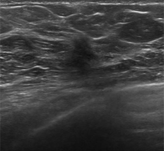
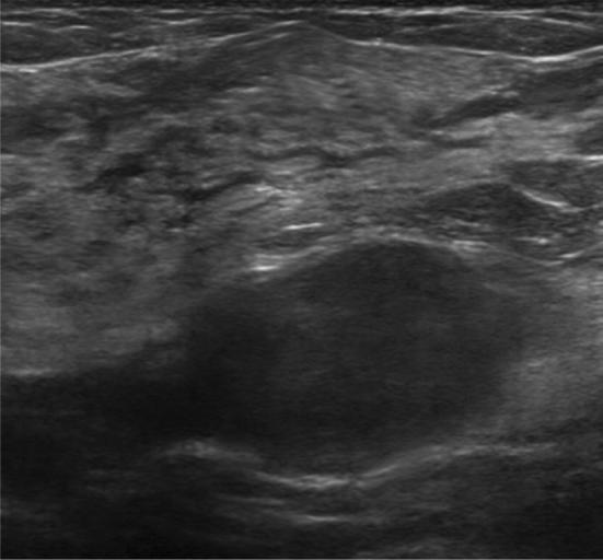
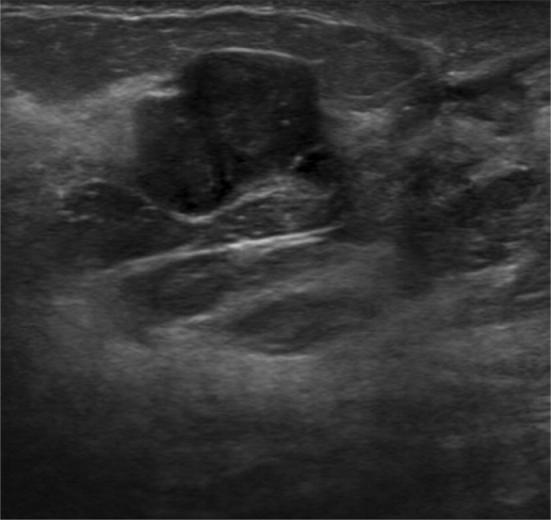
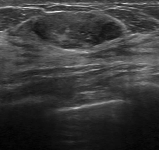
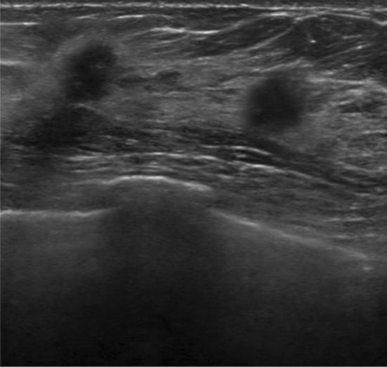
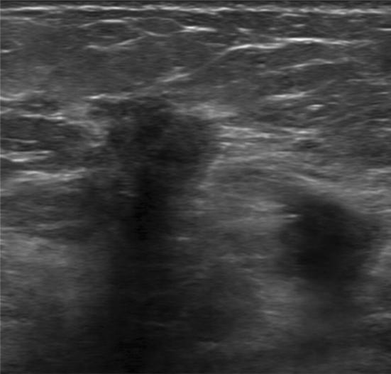
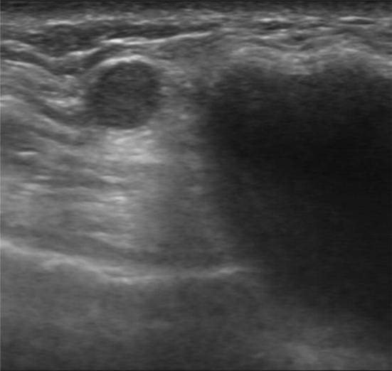
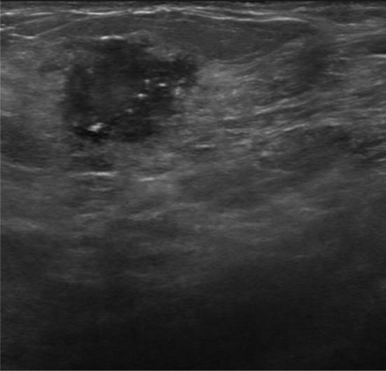
Methods: From June 2016 to November 2016, 155 patients with both benign and malignant breast lesions were selected from our institute to undergo US localization before surgery. The lesions included were: sonographically visible and nonpalpable lesions; palpable lesions for which a surgeon had requested US localization to better evaluate the site and extension; sonographically visible, multifocal breast lesions, both palpable and nonpalpable. US localization was performed using standard linear transducers (Siemens 18 L6, 5.5-8 MHz, 5.6 cm, ACUSON S2000 System, Siemens Medical Solutions). The radiologist used a skin pen to mark the site of the lesion, and the reported lesion's depth and distance from the nipple and pectoral muscle were recorded. The lesions were completely excised by a team of breast surgeons, and the surgical specimens were sent to the Radiology Department for radiological evaluation and to the Pathology Department for histological assessment.
Results: In 155 patients who underwent to preoperative US localization, 188 lesions were found, and the location of each lesion was marked with a skin pen. A total of 181 lesions were confirmed by the final histopathologic exam (96.28%); 132 of them (72.92%) were malignant, and 124 of these (93.93%) showed free margins.
Conclusions: US-guided preoperative localization of sonographically visible breast lesions is a simple and nontraumatic procedure with high specificity and is a useful tool for obtaining accurate surgical margins.
Obiettivo: Lo scopo dello studio è di verificare se la localizzazione preoperatoria eco-guidata delle lesioni mammarie sia una tecnica adeguata per una corretta e sicura resezione chirurgica ed è altresì quello di contribuire positivamente ed efficacemente, con i risultati ottenuti dal nostro istituto, all’approfondimento di questo argomento nella letteratura scientifica.
Metodi: Dal giugno 2016 al novembre 2016, 155 pazienti con lesioni mammarie benigne e maligne sono state selezionate dal nostro istituto per sostenere una localizzazione ecografica prima della seduta chirurgica.
Le lesioni considerate sono state:
lesioni ecograficamente visibili e non palpabili;
lesioni palpabili per le quali il chirurgo avesse richiesto una localizzazione ecografica per meglio valutarne sito ed estensione;
lesioni mammarie multifocali, ecograficamente visibili, sia palpabili che non palpabili.
La localizzazione ecografica è stata eseguita utilizzando trasduttori lineari standard (Siemens 18 L 6, 5.5–8 MHz, 5.6 cm, ACUSON S2000 System, Siemens Medical Solution).
Il radiologo ha utilizzato una penna per uso cutaneo per marcare il sito della lesione ed ha, dunque, calcolato e registrato profondità e distanza dal capezzolo e dal muscolo pettorale.
Le lesioni sono state completamente escisse dal team dei chirurghi mammari e i campioni chirurgici sono stati inviati al Dipartimento di Radiologia per una valutazione radiologica ed al Dipartimento di Anatomia patologica per una valutazione istologica.
Risultati: Nelle 155 pazienti, che sono state sottoposte a localizzazione ecografica pre-operatoria, sono state riscontrate 188 lesioni e il sito di ognuna di esse è stato marcato con una penna ad uso cutaneo.
Un totale di 181 lesioni è stato confermato dall’esame isto-patologico finale (96.28%); 132 di queste (72.92%) sono risultate maligne e 124 di queste ultime (93.93%) mostravano margini liberi da malattia.
Conclusioni: La localizzazione preoperatoria eco-guidata di lesioni mammarie ecograficamente visibili è una procedura semplice e non traumatica con un’elevata specificità ed è una metodica ideale per l’ottenimento di margini chirurgici indenni.
Keywords: Breast; Breast cancer; Histopathologic exam; Localization; Skin tattoo; Ultrasound.
Conflict of interest statement
Conflict of interest
The authors declare that they have no conflict of interest.
Ethical approval
All procedures performed in studies involving human participants were in accordance with the ethical standards of the institutional and/or national research committee and with the 1964 Helsinki declaration and its later amendments or comparable ethical standards.
Informed consent
For this type of study, formal consent is not required.
Figures
References
-
- Anderson BO, Lipscomb J, Murillo RH, et al. Disease Control Priorities, Third Edition (Volume 3) Washington (DC): The International Bank for Reconstruction and Development/The World Bank; 2015. - PubMed
-
- Krekel NM, Zonderhuis BM, Schreurs HW, et al. Ultrasound-guided breast-sparing surgery to improve cosmetic outcomes and quality of life. A prospective multicentrerandomised controlled clinical trial comparing ultrasound-guided surgery to traditional palpation-guided surgery (COBALT trial) BMC Surg. 2011;11:8. doi: 10.1186/1471-2482-11-8. - DOI - PMC - PubMed
MeSH terms
LinkOut - more resources
Full Text Sources
Medical
