

Sorafenib and docosahexaenoic acid act in synergy to suppress cancer cell viability: a role of heme oxygenase 1
- PMID: 30367621
- PMCID: PMC6204058
- DOI: 10.1186/s12885-018-4946-9
Sorafenib and docosahexaenoic acid act in synergy to suppress cancer cell viability: a role of heme oxygenase 1
Abstract
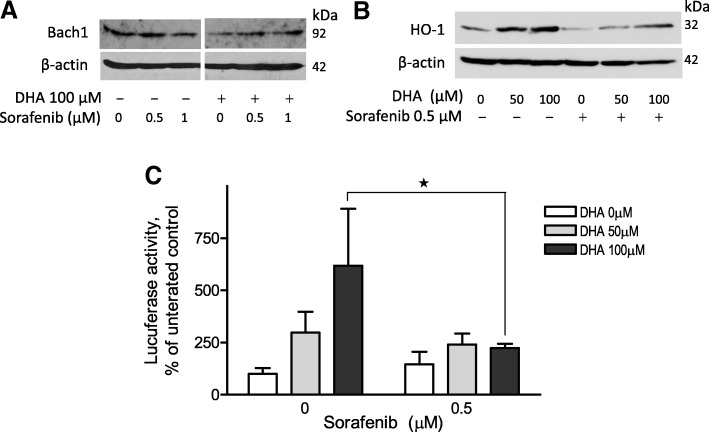
Background: Docosahexaenoic acid (DHA) is a long chain n-3 polyunsaturated fatty acid that has anticancer activity. Heme oxygenase 1 (HO-1) is a potential therapeutic target due to its cytoprotective activity in cancer cells. We recently reported that DHA induces HO-1 gene transcription in human cancer cells by augmenting the degradation of Bach1 protein, which functions as a negative regulator of HO-1. Since the degradation of Bach1 protein relies on protein phosphorylation, we hypothesized that DHA-induced HO-1 gene transcription could be attenuated by kinase inhibitors, resulting in an enhanced cytotoxicity. Sorafenib, a tyrosine kinase inhibitor, was first applied to test our hypothesis.
Methods: Human cancer cell lines and a xenograft nude mouse model were applied to test our hypothesis. Gene expression was analyzed by western blot analysis and reporter gene assay. Cell viability was analyzed using a colorimetric assay. Isobologram was applied to analyze drug action.
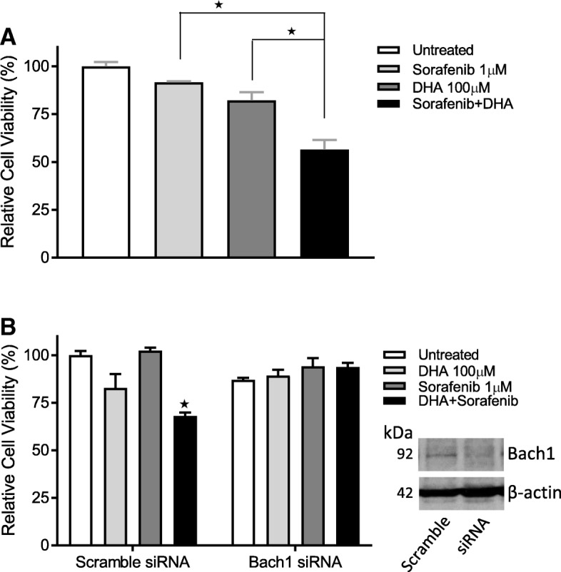
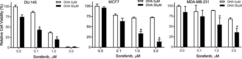
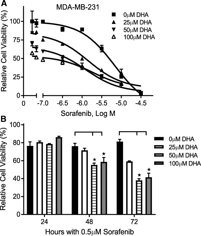
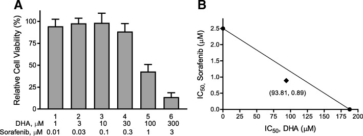
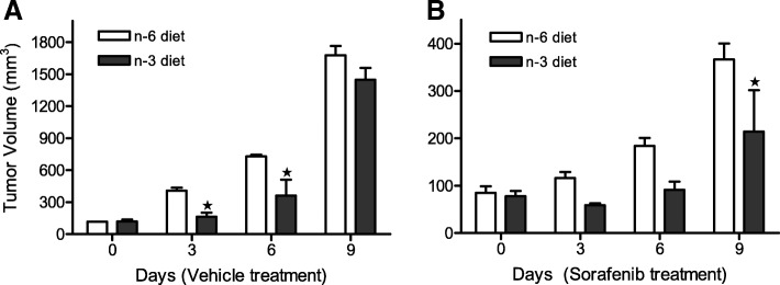
Results: Pretreatment of cancer cells with Sorafenib significantly attenuated DHA-induced degradation of Bach1 protein. Consequently, DHA-induced HO-1 gene transcription was reversed by Sorafenib as evidenced by western blot and reporter gene analysis. Sorafenib acted synergistically with DHA to suppress cancer cell viability in various human cancer cell lines and suppressed tumor xenograft growth in mice fed a fish oil enriched diet (high n-3/DHA), as compared to mice fed a corn oil (high n-6) diet. Screening of the NCI-Oncology Drug Set IV identified a group of anticancer compounds, including Sorafenib, which enhanced DHA's cytotoxicity, as well as a set of compounds that attenuated DHA's cytotoxicity.
Conclusions: We demonstrate that sorafenib attenuates DHA-induced HO-1 expression and acts in synergy with DHA to suppress cancer cell viability and tumor growth. Considering the known health benefits of DHA and the clinical effectiveness of Sorafenib, their combination is an attractive therapeutic strategy against cancer.
Keywords: Cancer; Docosahexaenoic acid; Heme oxygenase 1; Sorafenib; Synergy.
Conflict of interest statement
Ethics approval and consent to participate
Athymic nude mice (Foxn1nu) were purchased from Envigo (United Kingdom) and were used for in vivo evaluation of Sorafenib and DHA in accordance with the Institute Animal Care and Use Committee procedures and guidelines (Institute IACUC Protocol 100,861–14-025-SSH). All human cell lines were approved for use in this study by the Institutional Review Board (IRB protocol 4381).
Consent for publication
Not applicable.
Competing interest
The authors have no competing interest to disclose.
Publisher’s Note
Springer Nature remains neutral with regard to jurisdictional claims in published maps and institutional affiliations.
Figures
Similar articles
-
Characterization of docosahexaenoic acid (DHA)-induced heme oxygenase-1 (HO-1) expression in human cancer cells: the importance of enhanced BTB and CNC homology 1 (Bach1) degradation.J Nutr Biochem. 2014 May;25(5):515-25. doi: 10.1016/j.jnutbio.2013.12.011. Epub 2014 Feb 4. J Nutr Biochem. 2014. PMID: 24613086 Free PMC article.
-
Inhibition of matrix metalloproteinase-9 expression by docosahexaenoic acid mediated by heme oxygenase 1 in 12-O-tetradecanoylphorbol-13-acetate-induced MCF-7 human breast cancer cells.Arch Toxicol. 2013 May;87(5):857-69. doi: 10.1007/s00204-012-1003-3. Epub 2013 Jan 4. Arch Toxicol. 2013. PMID: 23288142
-
Docosahexaenoic acid inhibition of inflammation is partially via cross-talk between Nrf2/heme oxygenase 1 and IKK/NF-κB pathways.J Nutr Biochem. 2013 Jan;24(1):204-12. doi: 10.1016/j.jnutbio.2012.05.003. Epub 2012 Aug 15. J Nutr Biochem. 2013. PMID: 22901690
-
Docosahexaenoic acid: a natural powerful adjuvant that improves efficacy for anticancer treatment with no adverse effects.Biofactors. 2011 Nov-Dec;37(6):399-412. doi: 10.1002/biof.181. Epub 2011 Oct 28. Biofactors. 2011. PMID: 22038684 Review.
-
Docosahexaenoic Acid as Master Regulator of Cellular Antioxidant Defenses: A Systematic Review.Antioxidants (Basel). 2023 Jun 15;12(6):1283. doi: 10.3390/antiox12061283. Antioxidants (Basel). 2023. PMID: 37372014 Free PMC article. Review.
Cited by
-
The Potential of DHA as Cancer Therapy Strategies: A Narrative Review of In Vitro Cytotoxicity Trials.Nutrients. 2023 Apr 21;15(8):2006. doi: 10.3390/nu15082006. Nutrients. 2023. PMID: 37111226 Free PMC article. Review.
-
Cytoprotective Role of Heme Oxygenase-1 in Cancer Chemoresistance: Focus on Antioxidant, Antiapoptotic, and Pro-Autophagy Properties.Antioxidants (Basel). 2023 Jun 5;12(6):1217. doi: 10.3390/antiox12061217. Antioxidants (Basel). 2023. PMID: 37371947 Free PMC article. Review.
-
Nrf2 as a Potential Mediator of Cardiovascular Risk in Metabolic Diseases.Front Pharmacol. 2019 Apr 12;10:382. doi: 10.3389/fphar.2019.00382. eCollection 2019. Front Pharmacol. 2019. PMID: 31031630 Free PMC article. Review.
-
The Effects of Omega-3 Polyunsaturated Fatty Acids on Breast Cancer as a Preventive Measure or as an Adjunct to Conventional Treatments.Nutrients. 2023 Mar 7;15(6):1310. doi: 10.3390/nu15061310. Nutrients. 2023. PMID: 36986040 Free PMC article.
-
Docosahexaenoic Acid Coordinating with Sodium Selenite Promotes Paraptosis in Colorectal Cancer Cells by Disrupting the Redox Homeostasis and Activating the MAPK Pathway.Nutrients. 2024 Jun 1;16(11):1737. doi: 10.3390/nu16111737. Nutrients. 2024. PMID: 38892670 Free PMC article.
References
-
- Reddy BS, Burill C, Rigotty J. Effect of diets high in omega-3 and omega-6 fatty acids on initiation and postinitiation stages of colon carcinogenesis. Cancer Res. 1991;51(2):487–491. - PubMed
-
- Hirose M, Masuda A, Ito N, Kamano K, Okuyama H. Effects of dietary perilla oil, soybean oil and safflower oil on 7,12-dimethylbenz[a]anthracene (DMBA) and 1,2-dimethyl-hydrazine (DMH)-induced mammary gland and colon carcinogenesis in female SD rats. Carcinogenesis. 1990;11(5):731–735. doi: 10.1093/carcin/11.5.731. - DOI - PubMed
MeSH terms
Substances
Grants and funding
LinkOut - more resources
Full Text Sources
Research Materials
Miscellaneous
