. 2018 Oct 26;19(1):207.
doi: 10.1186/s12931-018-0915-8.

Selective activation and proliferation of a quiescent stem cell population in the neuroepithelial body microenvironment

Affiliations

Selective activation and proliferation of a quiescent stem cell population in the neuroepithelial body microenvironment

Line Verckist et al. Respir Res. .

Abstract

Background: The microenvironment (ME) of neuroepithelial bodies (NEBs) harbors densely innervated groups of pulmonary neuroendocrine cells that are covered by Clara-like cells (CLCs) and is believed to be important during development and for adult airway epithelial repair after severe injury. Yet, little is known about its potential stem cell characteristics in healthy postnatal lungs.

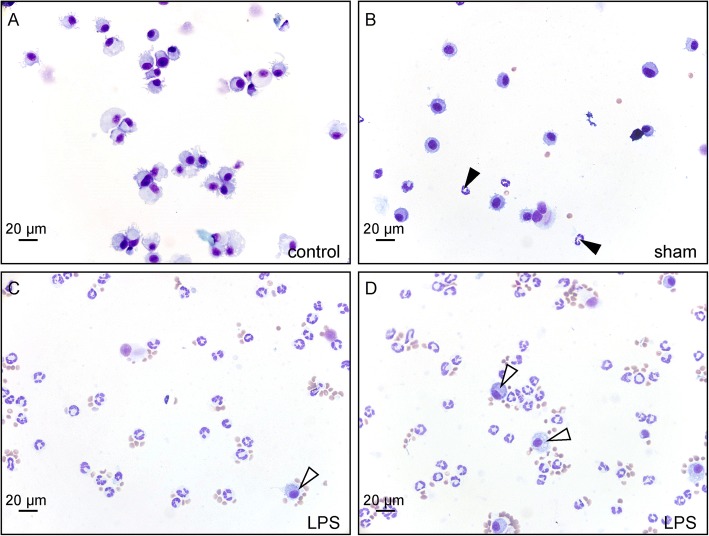
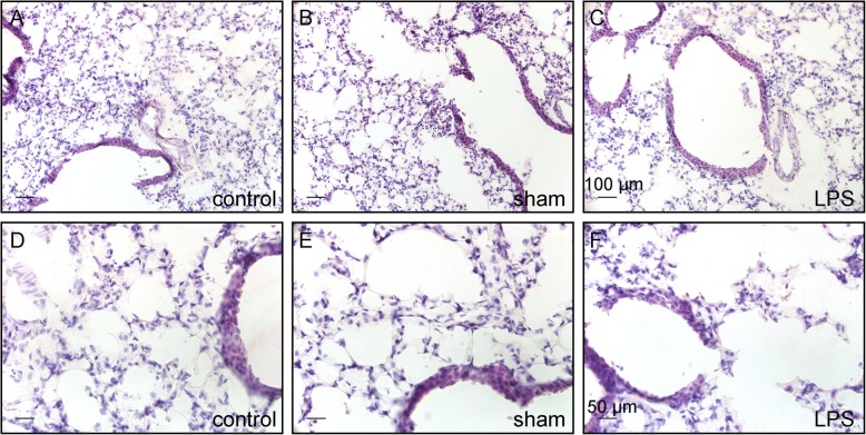
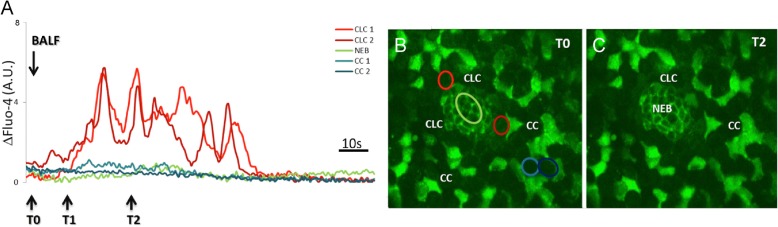
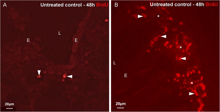
Methods: Transient mild lung inflammation was induced in mice via a single low-dose intratracheal instillation of lipopolysaccharide (LPS). Bronchoalveolar lavage fluid (BALF), collected 16 h after LPS instillation, was used to challenge the NEB ME in ex vivo lung slices of control mice. Proliferating cells in the NEB ME were identified and quantified following simultaneous LPS instillation and BrdU injection.

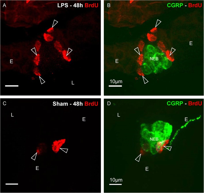
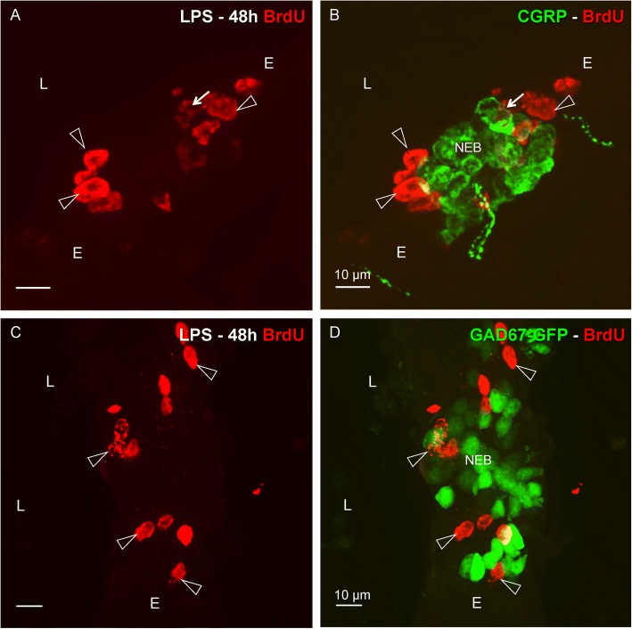
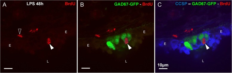
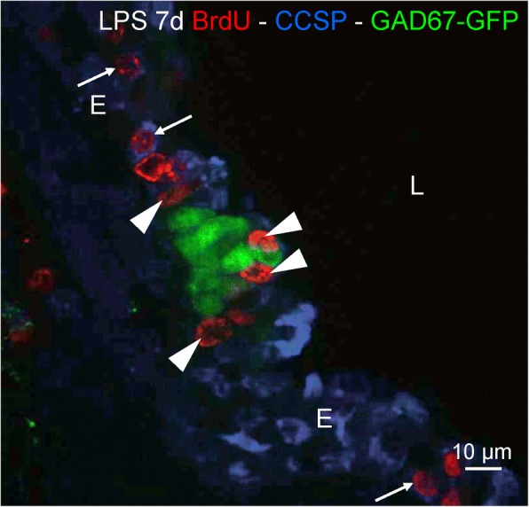
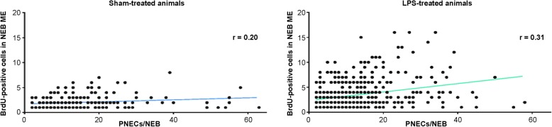
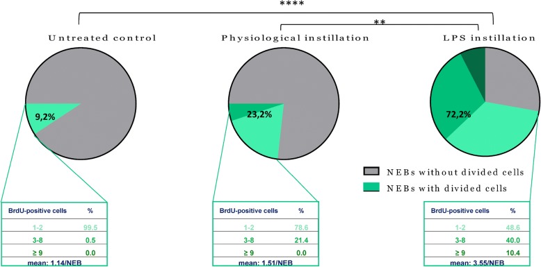
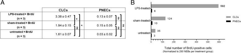
Results: The applied LPS protocol induced very mild and transient lung injury. Challenge of lung slices with BALF of LPS-treated mice resulted in selective Ca2+-mediated activation of CLCs in the NEB ME of control mice. Forty-eight hours after LPS challenge, a remarkably selective and significant increase in the number of divided (BrdU-labeled) cells surrounding NEBs was observed in lung sections of LPS-challenged mice. Proliferating cells were identified as CLCs.

Conclusions: A highly reproducible and minimally invasive lung inflammation model was validated for inducing selective activation of a quiescent stem cell population in the NEB ME. The model creates new opportunities for unraveling the cellular mechanisms/pathways regulating silencing, activation, proliferation and differentiation of this unique postnatal airway epithelial stem cell population.

Keywords: Airway epithelium; Clara-like cells; Lipopolysaccharide; Neuroepithelial body microenvironment; Proliferation; Pulmonary neuroendocrine cells; Stem cell niche.


Conflict of interest statement

Ethics approval

National and international principles of laboratory animal care were followed, and experiments were approved by the local animal ethics committee of the University of Antwerp (ECD 2014–66 and 2017–49).

Consent for publication

Not applicable.

Competing interests

The authors declare that they have no competing interests.

Publisher’s Note

Springer Nature remains neutral with regard to jurisdictional claims in published maps and institutional affiliations.

Figures

Fig. 1
Fig. 1
Representative images, with similar cell densities, of Diff-Quick-stained cytospin preparations of BALF collected from mice that received no instillation (untreated healthy control animal; a), 16 h after an intratracheal instillation with 0.9% NaCl (sham-treatment; b), or with LPS (c, d). Cell densities of the cytospin preparations were ‘normalized’ between the experimental groups and are therefore unrelated to the initial cell numbers in the BALF. a BALF of a healthy control mouse contains virtually no other cells than macrophages (acentric oval nuclei and bluish cytoplasm). b Some neutrophils (segmented nuclei and unstained cytoplasm; arrowheads) appear to be infiltrated in the airways after a sham instillation but macrophages still constitute the majority of cells in this sample. c, d Massive neutrophil influx is seen as a response to the LPS instillation, and consequently a relatively low number of macrophages (open arrowheads). Note that the preparations of treated animals also harbor some red blood cells (small brownish dots)
Fig. 2
Fig. 2
Representative images of HE-stained lung cryosections from untreated control (a, d), and 48 h after intratracheal instillation with 0.9% NaCl (sham-treatment; b, e), or with LPS (c, f). Note that lung morphology shows no clear histological differences between the groups
Fig. 3
Fig. 3
LCI representation of the effects of application of BALF of an LPS-challenged mouse to the NEB ME in a vibratome cut live lung slice of a control mouse. a Graphs plotting the time course of Fluo-4 fluorescence intensity, as an indicator for [Ca2+]i, in different cell types in the airway epithelium. To facilitate interpretation, grey levels of each region of interest (ROI) were adjusted for the basal level of (background) fluorescence at the start of imaging. After application of BALF (=T0), CLCs that surround the NEB cells show a calcium-mediated activation within about 10 s (=T1). The oscillating [Ca2+]i rise in CLCs continues (=T2) for more than a minute post-exposure. No changes in [Ca2+]i can be observed in NEB cells or CCs. b, c Corresponding pseudo-color time-lapse images of Fluo-4 fluorescence at two time points (B = T0; C = T2) after the application of BALF. Note that the ROIs corresponding to the graphs in (a) are represented in the same color code in image b
Fig. 4
Fig. 4
Immunostaining for BrdU (red Cy3 fluorescence) in cryosections of an intrapulmonary airway (a) and small intestine (b) of the same mouse that received no treatment, except for the BrdU injections (i.p.), 48 h and 24 h prior to sacrifice. a The airway epithelium rarely harbors divided BrdU-positive cells. The majority of BrdU-labeled nuclei (arrowheads) are located in subepithelial layers. b In the crypts (asterisks) and the basal parts of the villus epithelium of the small intestine, a large number of BrdU-labeled nuclei (arrowheads) –– i.e., originating from cells that have divided during the 48 h experimental window –– can be observed. L: airway lumen, E: airway epithelium
Fig. 5
Fig. 5
Immunostaining for BrdU (red Cy3 fluorescence) and CGRP (green FITC fluorescence) in intrapulmonary airways 48 h after an intratracheal LPS (a, b) or sham instillation (c, d) in WT-Bl6 mice. a After LPS challenge, clustered BrdU-positive (divided) cells are observed in the epithelial layer (open arrowheads). b Additional CGRP immunostaining reveals that the intraepithelial BrdU-labeled cells are typically grouped around NEBs. c, d After a sham instillation, intraepithelial BrdU-positive cells also appear to be located in the neighborhood of NEBs (open arrowhead), but are less numerous than after LPS treatment. L: airway lumen, E: airway epithelium
Fig. 6
Fig. 6
Comparison of the distribution of BrdU-labeled (red Cy3 fluorescence) airway epithelial cells 48 h after LPS challenge of WT-Bl6 (a, b) and GAD67-GFP mice (c, d). a, c Clustered BrdU-positive nuclei (open arrowheads) can be observed at distinct locations in the airway epithelium. Combination with CGRP immunostaining (b; green FITC fluorescence) for WT-Bl6 mice, or visualization of GFP-fluorescent NEB cells (d) in GAD67-GFP mice, reveals that the majority of divided cells that have incorporated BrdU are found in the immediate neighborhood of NEBs. No obvious differences can be seen between WT-Bl6 and GAD67-GFP mice. Note that only very occasionally, a BrdU-positive nucleus can be seen in a NEB cell (arrow; a, b). L: airway lumen, E: airway epithelium
Fig. 7
Fig. 7
Single confocal optical section of the airway epithelium in a cryosection of the lungs of a GAD67-GFP mouse 48 h after challenge with LPS. a BrdU-positive nucleus (arrowhead; red Cy3 fluorescence) in the airway epithelium, and some subepithelial BrdU staining (open arrowhead). b Combination with GAD67-GFP fluorescence (green), marking PNECs in the NEB ME. c Image of the three channels. In the same section, CCs/CLCs are immunostained for the Clara cell-specific protein CCSP (blue pseudo-color of Cy5 fluorescence), and are seen to surround GFP-labeled NEB cells. Note that the divided epithelial cell (BrdU-labeled nucleus; arrowhead) is located adjacent to the PNECs and co-stained with CCSP, and can therefore be identified as a CLC. L: airway lumen, E: airway epithelium
Fig. 8
Fig. 8
Distribution of divided (BrdU-stained; red Cy3 fluorescence) airway epithelial cells seven days after LPS challenge. Similar to the 48 h time window, several CCSP-immunostained (blue pseudo-color of Cy5 fluorescence), BrdU-positive nuclei (arrowheads) can be observed specifically surrounding GAD67-GFP fluorescent (green) PNECs in the NEB ME. Note, however, the additional presence of CCSP-stained BrdU-labeled CCs in the surrounding airway epithelium (open arrowheads). L: airway lumen, E: airway epithelium
Fig. 9
Fig. 9
Graph plotting the number of BrdU-positive (divided) cells per NEB ME as a function of the number of PNECs for each NEB that harbors BrdU-positive cells, both in sham- (left graph) and LPS-treated (right graph) mice (n = 10; 5 WT-Bl6 and 5 GAD67-GFP mice for each treatment group). The full data sets show no significant positive correlation between the size of NEBs and the number of BrdU-positive cells in their ME. r = Spearman correlation coefficient
Fig. 10
Fig. 10
Percentage of NEBs with cells in their ME that have divided during the 48 h experimental window. In the untreated controls (n = 5), less than 10% of the NEB MEs harbor divided cells. Interestingly, both the percentage of NEBs with BrdU-positive cells (about 23%) and the number of divided cells per NEB ME is higher in sham-treated mice (n = 10) than in untreated control mice. After LPS instillation (n = 10), however, about 72% of the NEBs show divided cells in their ME. The green part of the pie charts represents the percentage of NEBs with BrdU-positive cells, and is further subdivided based on the number of BrdU-labeled cells per NEB. The data in the framed areas represent the percentages of NEBs with one or two, three to eight, or nine and more BrdU-positive cells. The mean number of BrdU-positive cells is significantly different (Dunn’s multiple comparisons test) between the untreated control and the LPS-treated group (****, p < 0.0001) and between the sham- and LPS-treated group (**, p < 0.01)
Fig. 11
Fig. 11
Table providing the mean numbers of divided (BrdU-labeled) CLCs and PNECs per activated NEB ME (a) and graph representing the total numbers of BrdU-positive CLCs and PNECs (b) for all quantified NEBs in the three experimental groups (n = 3 GAD67-GFP mice per group)

Similar articles

Cited by

References

    1. Thurlbeck WM. Postnatal growth and development of the lung. Am Rev Respir Dis. 1975;111:803–844. - PubMed
    1. Giangreco A, Arwert EN, Rosewell IR, Snyder J, Watt FM, Stripp BR. Stem cells are dispensable for lung homeostasis but restore airways after injury. Proc Natl Acad Sci U S A. 2009;106:9286–9291. doi: 10.1073/pnas.0900668106. - DOI - PMC - PubMed
    1. Bertoncello I, McQualter JL. Lung stem cells: do they exist? Respirology. 2013;18:587–595. doi: 10.1111/resp.12073. - DOI - PubMed
    1. Stabler CT, Morrisey EE. Developmental pathways in lung regeneration. Cell Tissue Res. 2017;367:677–685. doi: 10.1007/s00441-016-2537-0. - DOI - PMC - PubMed
    1. Bishop AE. Pulmonary epithelial stem cells. Cell Prolif. 2004;37:89–96. doi: 10.1111/j.1365-2184.2004.00302.x. - DOI - PMC - PubMed