

Relevance of interferon-gamma in pathogenesis of life-threatening rapidly progressive interstitial lung disease in patients with dermatomyositis
- PMID: 30367666
- PMCID: PMC6235206
- DOI: 10.1186/s13075-018-1737-2
Relevance of interferon-gamma in pathogenesis of life-threatening rapidly progressive interstitial lung disease in patients with dermatomyositis
Abstract
Background: Dermatomyositis (DM) with rapidly progressive interstitial lung disease (DM RP-ILD) is a life-threatening condition. Serum cytokine levels are potentially suitable biomarkers for DM RP-ILD. However, the relationships among cytokine levels, lung imaging findings, and lung pathology have not been investigated. The aim of the present retrospective study was to determine the association between hypercytokinemia and lung inflammation in patients with DM RP-ILD.
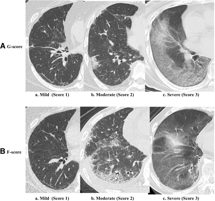
Methods: The study subjects were nine patients with life-threatening DM RP-ILD and severe hypoxemia (partial arterial oxygen pressure (PaO2)/fraction of inspired oxygen (FiO2) ratio ≤ 200) before receiving intensive care management, who were admitted to our hospital between 2006 and 2015. The controls included 10 patients with DM without RP-ILD and 19 healthy subjects. We assessed the association between serum cytokine levels and computed tomography (CT) scores of the lung (ground glass opacity-score, G-score; fibrosis-score, F-score). Lung, hilar lymph nodes, and spleen from two autopsies were examined by hematoxylin-eosin (H&E) staining and immunostaining.
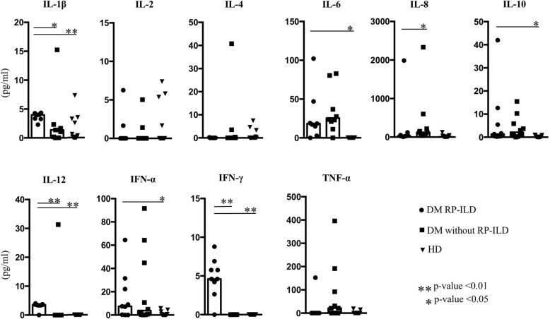
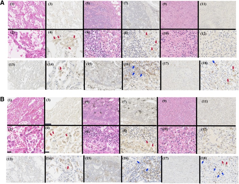
Results: Serum interferon (IFN)-γ, interleukin (IL)-1β and IL-12 levels were significantly higher in patients with DM RP-ILD than in the other two groups, whereas serum IL-6 levels were elevated in the two patient groups but not in the healthy subjects. Serum levels of IL-2, IL-4, IL-8, IL-10, IFN-α, and TNF (tumor necrosis factor)-α were not characteristically elevated in the DM RP-ILD group. Serum IFN-γ levels correlated with G-scores in patients with DM RP-ILD, while IL-1β was negatively correlation with F-scores. Immunohistochemical staining showed infiltration of numerous IFN-γ-positive histiocytes in the lung and hilar lymph nodes; but not in the spleen. Serum IL-6 levels did not correlate with the CT scores. Numerous IL-6-positive plasma cells were found in hilar lymph nodes, but not in the lungs or spleen.
Conclusions: Our results suggest strong IFN-γ-related immune reaction in the lungs and hilar lymph nodes of patients with life-threatening DM RP-ILD, and potential IFN-γ involvement in the pathogenesis of DM, specifically in the pulmonary lesions of RP-ILD.
Keywords: Dermatomyositis; IFN-γ; Rapidly progressive interstitial lung disease.
Conflict of interest statement
Ethics approval and consent to participate
Ethical approval was obtained from the University of Occupational and Environmental Health, Japan Ethics Committee. This retrospective study was approved by the institutional review board, and the requirement to obtain informed consent was waived.
Consent for publication
Not applicable.
Competing interests
Kei Sakata is an employee of Mitsubishi Tanabe Pharma. Shingo Nakayamada has received speaking fees from Bristol-Myers, Sanofi, AbbVie, Eisai, Eli Lilly, Chugai, Pfizer, Takeda (less than US $10,000 each), and research grants from Mitsubishi-Tanabe, Novartis and MSD. Dr Tanaka has received consulting fees, speaking fees, and/or honoraria from Mitsubishi Tanabe Pharma, Eisai, Pfizer, Abbott Immunology, Janssen, Takeda Industrial Pharma, Santen, AstraZeneca, Astellas, Asahi Kasei, UCB, and GlaxoSmithKline (less than US $10,000 each) and from AbbVie and Chugai (more than US $10,000 each) and research grants from Bristol-Myers Squibb, Mitsubishi Tanabe, MSD, Takeda Industrial Pharma, Astellas, Eisai, Chugai, Pfizer, and Daiichi-Sankyo. All other authors declare no conflict of interest.
Publisher’s Note
Springer Nature remains neutral with regard to jurisdictional claims in published maps and institutional affiliations.
Figures
References
Publication types
MeSH terms
Substances
LinkOut - more resources
Full Text Sources
Medical
Molecular Biology Databases
