Review
. 2019 May 1:319:7-15.
doi: 10.1016/j.jneumeth.2018.10.032. Epub 2018 Oct 24.

High resolution approaches for the identification of amyloid fragments in brain

Affiliations
Review

High resolution approaches for the identification of amyloid fragments in brain

J A Ross et al. J Neurosci Methods. .

Abstract

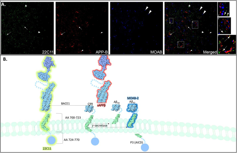
Background: It is now widely recognized that endogenous, picomolar concentrations of the 42 amino acid long peptide, amyloid-β (Aβ42) is secreted under normal physiological conditions and exerts important functional activity throughout neuronal intracellular compartments. Transgenic animal models that overexpress Aβ42 and its precursor, amyloid precursor protein (APP), have not provided predictive value in testing new treatments for Alzheimer's disease (AD), resulting in failed clinical trials. While these results are discouraging, they underscore the need to understand the physiological roles of Aβ42 and APP under normal conditions as well as at early pre- symptomatic stages of AD. New method: We describe the use of acrolein-perfusion in immunoelectron microscopy in combination with novel antibodies directed against endogenous murine Aβ42 and APP fragments to study abnormalities in the endolysosomal system at early stages of disease. The specific requirements, limitations and advantages of novel antibodies directed against human and murine Aβ42, APP and APP fragments are discussed as well as parameters for ultrastructural analysis of endolysosomal compartments.

Results: Novel antibodies and a detailed protocol for immunoelectron microscopy using acrolein as a fixative are described. Acrolein is shown to preserve intraneuronal Aβ42 species, as opposed to paraformaldehyde fixed tissue, which primarily preserves membrane bound species. Comparison with existing method(s): Technology sensitive enough to detect endogenous Aβ42 under physiological conditions has not been widely available. We describe a number of novel and highly sensitive antibodies have recently been developed that may facilitate the analysis of endogenous Aβ42.

Conclusions: Using novel and highly specific antibodies in combination with electron microscopy may reveal important information about the timing of aberrant protein accumulation, as well as the progression of abnormalities in the endolysosomal systems that sort and clear these peptides.

Keywords: Amyloid; Antibodies; Electron microscopy.


Figures

Figure 1:
Figure 1:. APP Fragments and Specific Antibody Recognition Sites.
The proteolytic processing of APP generally occurs via two divergent pathways. The non-amyloidogenic pathway is the most common route of processing in most cells, and results from cleavage of APP by α-secretases embedded in the plasma membrane. This cleavage results in the formation of sAPPα composed of amino acids 18–687 and a carboxyl terminal C83 fragment (α-CTF) composed of amino acids 688–770. The amyloidogenic pathway accounts for a smaller portion of APP processing. In this pathway, APP undergoes proteolytic cleavage by the aspartic protease β-secretase (BACE-1), which cuts APP on the luminal side of the membrane, releasing a soluble APPβ fragment (sAPPβ) composed of amino acids 18–671, and carboxyl terminal C99 fragment (β-CTF) composed of amino acids 672–770 (Vassar, Bennett et al. 1999). BACE-1 cleavage results in the formation of a new N-terminus with the first aspartic amino acid 672 of Aβ (LaFerla, Green et al. 2007), which is a neo-epitope detected by some antibodies. The C1/61 antibody binds the C-terminus of APP, thus detecting all full-length APP and related CTFs. Following BACE-1 cleavage, antibody JRF/N25 detects the C99 β-CTF. Subsequent cleavage of this β-CTF, at 38–43 amino acids downstream of this β-cleavage site by the γ-secretase, results in the release of the Aβ40,42, and to a lesser extent other Aβ peptides. To readily distinguish fragments Aβ40 and Aβ42, the N-terminal specific antibodies JRF/cAβ40/10 and JRF/cAβ 42/26 may be used, respectively. The structure of the APP molecule is attributed to David S. Goodsell and the RCSB Protein Data Bank (PDB), and was modified to depict fragments following proteolytic cleavage (D. Goodsell 2015).
Figure 2.
Figure 2.. Distinguished labeling patterns of 22C11, APP-β, and MOAB-2 antibodies in the naïve rat.
The epitopes labeled by three different antibodies within the APP protein readily distinguish three distinct fragments with unique distributions. The 22C11 (green) antibody labels an epitope that reveals full length APP, while the APP-β (red) antibody labels sAPP-β, the fragment resulting from BACE-1 cleavage of full-length APP, and MOAB-2 (blue) an antibody that labels the intracellular fragment that results from γ-secretase cleavage of the sAPP-β fragment. can be readily distinguished using low-resolution immunofluorescence techniques. Individually labeled puncta in close proximity (panel a,a’) may represent full length, membrane-bound APP and highlights the distinct region of the APP peptide in which the antibody-specific epitope is present; for example, 22C11 labeling (green) is the N-terminal extracellular region of APP, while MOAB-2 (blue) embedded within the membrane, and APP-β (red) that labels the C-terminus is facing the cytoplasmic side of the plasma membrane. There are several occurrences of co-localization, which primarily occur between the 22C11 and APP-β antibodies (yellow puncta), that indicate β-CTF labeling. Importantly, there are few occasions in which MOAB-2 labeling is co-localized with APP-β (magenta), which may be readily distinguished from γ-secretase cleavage products identified by MOAB-2 labeling alone. There were no occurrences of MOAB-2 immunoreactivity with 22C11 labeling. MOAB-2 is also more frequently visualized as individual puncta, and further away from 22C11 and APP-β immunoreactivity, alluding to the putative intracellular localization of Aβ42 peptides. However, it should be noted that electron microscopy is necessary to define the subcellular localization of these fragments with any certainty.
Figure 3:
Figure 3:. APP Processing and the Endolysosomal System.
The fate of APP is dictated largely by its subcellular localization, thus highlighting the importance of trafficking in parallel with proteolytic cleavage. Transmembrane proteins such as APP that are targeted for degradation enter the endosomal-lysosomal pathway by undergoing endocytosis, autophagy or phagocytosis. APP is internalized from the plasma membrane via endocytosis and further processed in endocytic, recycling and lysosomal compartments. In addition, once in the endosome, APP may be transported back to the TGN (G, blue) via retromer proteins (Vieira, Rebelo et al. 2010), following recognition by the sortilin related receptor (SORLA). Thus, transference of various forms of APP and its fragments occurs via the highly dynamic membrane enclosed vesicular structures that are compositionally and functionally distinct. These structures have been well characterized and include the early endosome, recycling endosome, late endosome (End, green) and lysosome (Lys, red) (Huotari and Helenius 2011). Arrow heads point to immunogold labeled Aβ42.
Figure 4:
Figure 4:. Electron microscopy.
Following immunohistochemical procedures, tissues are prepared for visualization under the electron microscope with osmification, serial dehydration, flat-embedding, and tissue sectioning at 74 nm on an ultramicrotome (Commons, Beck et al. 2001). Sections are collected on copper mesh grids and examined using an electron microscope (Morgani, Fei Company, Hillsboro, OR). Digital images are viewed and captured using the AMT advantage HR HR-B CCD camera system (Advance Microscopy Techniques, Danvers, MA). Electron micrograph images are then prepared using Adobe Photoshop to adjust the brightness and contrast.
Figure 5.
Figure 5.. Aβ42 subcellular localization.
A. Immunoelectron micrographs of TH-immunoreactive dendrites (TH-d), one of which is dually labeled with immunogold Aβ42 (arrow heads). More specifically, Aβ42 is localized to a lysosomal (Lys) compartment within the dendrite, identified at the ultrastructural level. B. Example of an autolysosome that contains heterogeneous mixture of electron dense materials, including immunogold labeled Aβ42. C. Immunogold labeled Aβ42 is localized to axon terminals (at) presynaptic to TH immunolabeled dendrite. Here, immunogold labeled Aβ42 is associated with mitochondrial membranes (m). D. Immunoelectron micrograph of immunogold labeled Aβ42 localized to an axon terminal filled with dense core vesicles (dcv), a subcellular compartment derived from multivesicular bodies that frequently contain neuropeptides co-packaged with fast acting neurotransmitters that may be released from asynaptic sites. E. TH-immunolabeled cell body that contains several lysosomes with immunogold labeled Aβ42; immunogold labeled Aβ42 is also present on the cell surface, potentially indicating secretion into the extracellular space.

Similar articles

Cited by

References

    1. Bainton DF (1981). “The discovery of lysosomes.” J Cell Biol 91(3 Pt 2): 66s–76s. - PMC - PubMed
    1. Barnham KJ, McKinstry WJ, Multhaup G, Galatis D, Morton CJ, Curtain CC, Williamson NA, White AR, Hinds MG, Norton RS, Beyreuther K, Masters CL, Parker MW and Cappai R (2003). “Structure of the Alzheimer’s disease amyloid precursor protein copper binding domain. A regulator of neuronal copper homeostasis.” J Biol Chem 278(19): 17401–17407. - PubMed
    1. Burgos PV, Mardones GA, Rojas AL, daSilva LL, Prabhu Y, Hurley JH and Bonifacino JS (2010). “Sorting of the Alzheimer’s disease amyloid precursor protein mediated by the AP-4 complex.” Dev Cell 18(3): 425–436. - PMC - PubMed
    1. Choi JH, Berger JD, Mazzella MJ, Morales-Corraliza J, Cataldo AM, Nixon RA, Ginsberg SD, Levy E and Mathews PM (2009). “Age-dependent dysregulation of brain amyloid precursor protein in the Ts65Dn Down syndrome mouse model.” J Neurochem 110(6): 1818–1827. - PMC - PubMed
    1. Cirrito JR, Kang JE, Lee J, Stewart FR, Verges DK, Silverio LM, Bu G, Mennerick S and Holtzman DM (2008). “Endocytosis is required for synaptic activity-dependent release of amyloid-beta in vivo.” Neuron 58(1): 42–51. - PMC - PubMed

Publication types

LinkOut - more resources