

Protective effects of mangafodipir against chemotherapy-induced ovarian damage in mice
- PMID: 30368246
- PMCID: PMC6204278
- DOI: 10.1186/s12958-018-0426-y
Protective effects of mangafodipir against chemotherapy-induced ovarian damage in mice
Abstract
Background: Given the seriousness of chemotherapy-induced ovarian injury in female cancer patients, the preservation of fertility, including through the use of cryopreservation technology and pharmaceuticals, requires investigation. Previous studies have shown that damage to the ovaries is related to oxidative stress caused by anticancer drugs. Therefore, superoxide dismutase (SOD) may represent a key factor in the pharmacological protection of the ovaries. The aim of our study was to identify the effects of mangafodipir, a manganese chelate and SOD-mimetic, on suppression of apoptosis in granulosa cells and primordial follicle activation induced by anticancer drugs.
Methods: Cell viability assays using methyltrichlorosilane solutions and immunoblotting for cleaved caspase-3 were performed in in vitro experiments with the simultaneous addition of mangafodipir to human non-luteinized granulosa cell line (HGrC) cultures treated with hydrogen peroxide (H2O2), cisplatin, or paclitaxel. Count and morphological analyses of follicles at each developing stage in the ovaries and immunohistochemistry for cleaved caspase-3, Ki67 and 4-hydroxynonenal, a marker for oxidative stress, were also performed using mangafodipir-injected 6-week-old female ICR mice treated with cisplatin or paclitaxel. Further, mangafodipir was injected into 6-week-old female BALB/c mice inoculated with ES-2 to analyze whether mangafodipir inhibits the anti-tumor effects of cisplatin or paclitaxel treatment.
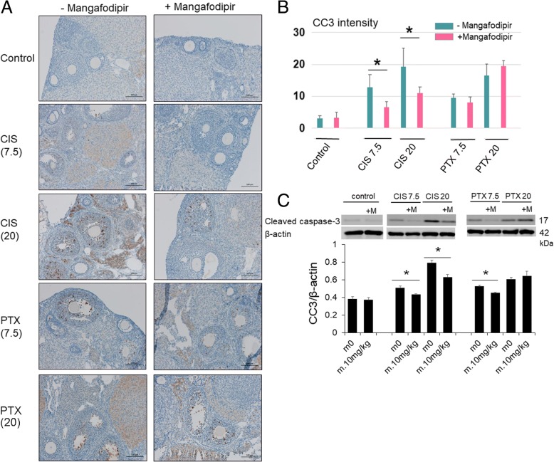
Results: Mangafodipir attenuated apoptosis induced by H2O2 and anticancer drugs in vitro. Mangafodipir also decreased the expression of 4-hydroxynonenal and reduced cisplatin- and paclitaxel-induced apoptosis in granulosa cells in vivo. In addition, mangafodipir inhibited the loss of primordial follicles. Tumor xenograft studies in mice showed that mangafodipir did not affect anticancer drug antitumor effects.
Conclusions: Oxidative stress might be one of the mechanisms of cisplatin- and paclitaxel-induced the loss of primordial follicles. Mangafodipir can reduce cisplatin- and paclitaxel-induced apoptosis in granulosa cells and primordial follicle activation partially via its SOD activity. At the same time, mangafodipir might have other potential mechanisms to inhibit the activation of primordial follicles. Further, mangafodipir attenuated the ovarian damage caused by cisplatin and paclitaxel without affecting their antitumor activities. Mangafodipir, therefore, though its efficacy might be limited, may be a new option for the preservation of fertility during anticancer treatment.
Keywords: Anticancer drug; Follicle; Mangafodipir; Ovary; Oxidative stress.
Conflict of interest statement
Ethics approval and consent to participate
The study was approved by the Division of Experimental Animals at Nagoya University Graduate School of Medicine.
Consent for publication
Not applicable.
Competing interests
The authors declare no potential conflicts of interest with respect to the research, authorship, and/or publication of this article.
Publisher’s Note
Springer Nature remains neutral with regard to jurisdictional claims in published maps and institutional affiliations.
Figures


Similar articles
-
Improvement of the therapeutic index of anticancer drugs by the superoxide dismutase mimic mangafodipir.J Natl Cancer Inst. 2006 Feb 15;98(4):236-44. doi: 10.1093/jnci/djj049. J Natl Cancer Inst. 2006. PMID: 16478742
-
The magnitude of gonadotoxicity of chemotherapy drugs on ovarian follicles and granulosa cells varies depending upon the category of the drugs and the type of granulosa cells.Hum Reprod. 2015 Dec;30(12):2926-35. doi: 10.1093/humrep/dev256. Epub 2015 Oct 13. Hum Reprod. 2015. PMID: 26466914
-
Ceramide-1-phosphate has protective properties against cyclophosphamide-induced ovarian damage in a mice model of premature ovarian failure.Hum Reprod. 2018 May 1;33(5):844-859. doi: 10.1093/humrep/dey045. Hum Reprod. 2018. PMID: 29534229
-
Prevention of chemotherapy-induced ovarian damage.Fertil Steril. 2016 Jan;105(1):20-9. doi: 10.1016/j.fertnstert.2015.11.043. Epub 2015 Dec 8. Fertil Steril. 2016. PMID: 26677788 Review.
-
Ovarian microenvironment: challenges and opportunities in protecting against chemotherapy-associated ovarian damage.Hum Reprod Update. 2024 Oct 1;30(5):614-647. doi: 10.1093/humupd/dmae020. Hum Reprod Update. 2024. PMID: 38942605 Free PMC article. Review.
Cited by
-
Inhibition of Drp1 orchestrates the responsiveness of breast cancer cells to paclitaxel but insignificantly relieves paclitaxel-related ovarian damage in mice.Sci Rep. 2023 Dec 20;13(1):22782. doi: 10.1038/s41598-023-49578-0. Sci Rep. 2023. PMID: 38129495 Free PMC article.
-
Novel ovarian endometriosis model causes infertility via iron-mediated oxidative stress in mice.Redox Biol. 2020 Oct;37:101726. doi: 10.1016/j.redox.2020.101726. Epub 2020 Sep 15. Redox Biol. 2020. PMID: 32961443 Free PMC article.
-
The role of oxidative stress in ovarian aging: a review.J Ovarian Res. 2022 Sep 1;15(1):100. doi: 10.1186/s13048-022-01032-x. J Ovarian Res. 2022. PMID: 36050696 Free PMC article. Review.
-
Transcriptomic Analysis of circRNAs and mRNAs Reveals a Complex Regulatory Network That Participate in Follicular Development in Chickens.Front Genet. 2020 May 15;11:503. doi: 10.3389/fgene.2020.00503. eCollection 2020. Front Genet. 2020. PMID: 32499821 Free PMC article.
-
Can Some Anticancer Treatments Preserve the Ovarian Reserve?Oncologist. 2021 Jun;26(6):492-503. doi: 10.1002/onco.13675. Epub 2021 Jan 29. Oncologist. 2021. PMID: 33458904 Free PMC article. Review.
References
-
- Loren AW, Mangu PB, Beck LN, Brennan L, Magdalinski AJ, Partridge AH, Quinn G, Wallace WH, Oktay K. American Society of Clinical O. fertility preservation for patients with cancer: American Society of Clinical Oncology clinical practice guideline update. J Clin Oncol. 2013;31:2500–2510. doi: 10.1200/JCO.2013.49.2678. - DOI - PMC - PubMed
-
- Fisch B, Abir R. Female fertility preservation: past. present and future Reproduction. 2018;156:F11–F27. - PubMed
-
- Murase T, Iwase A, Komatsu K, Bayasula NT, Osuka S, Takikawa S, Goto M, Kotani T, Kikkawa F. Follicle dynamics: visualization and analysis of follicle growth and maturation using murine ovarian tissue culture. J Assist Reprod Genet. 2018;35:339–343. doi: 10.1007/s10815-017-1073-5. - DOI - PMC - PubMed
MeSH terms
Substances
Grants and funding
LinkOut - more resources
Full Text Sources
Research Materials
