. 2019 Jan;76(2):381-395.
doi: 10.1007/s00018-018-2957-y. Epub 2018 Oct 27.

NudCL2 is an Hsp90 cochaperone to regulate sister chromatid cohesion by stabilizing cohesin subunits

Affiliations

NudCL2 is an Hsp90 cochaperone to regulate sister chromatid cohesion by stabilizing cohesin subunits

Yuehong Yang et al. Cell Mol Life Sci. 2019 Jan.

Abstract

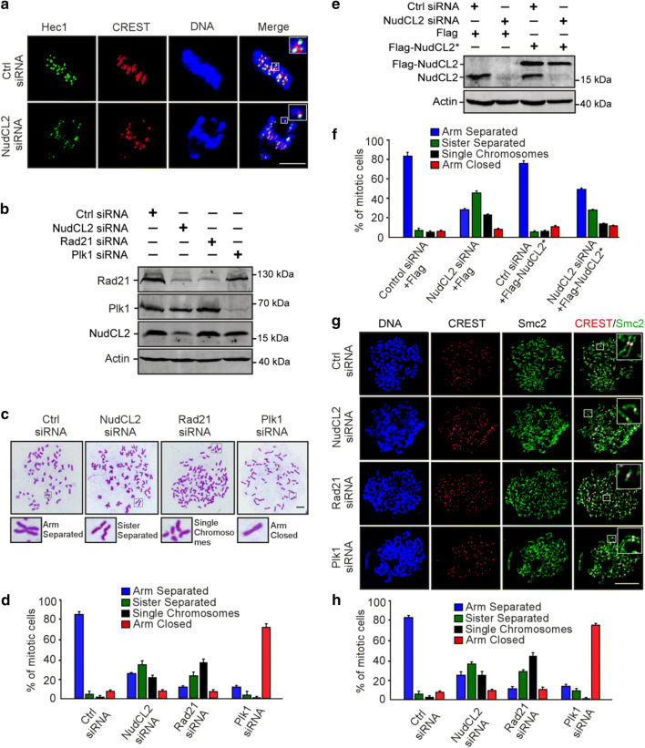
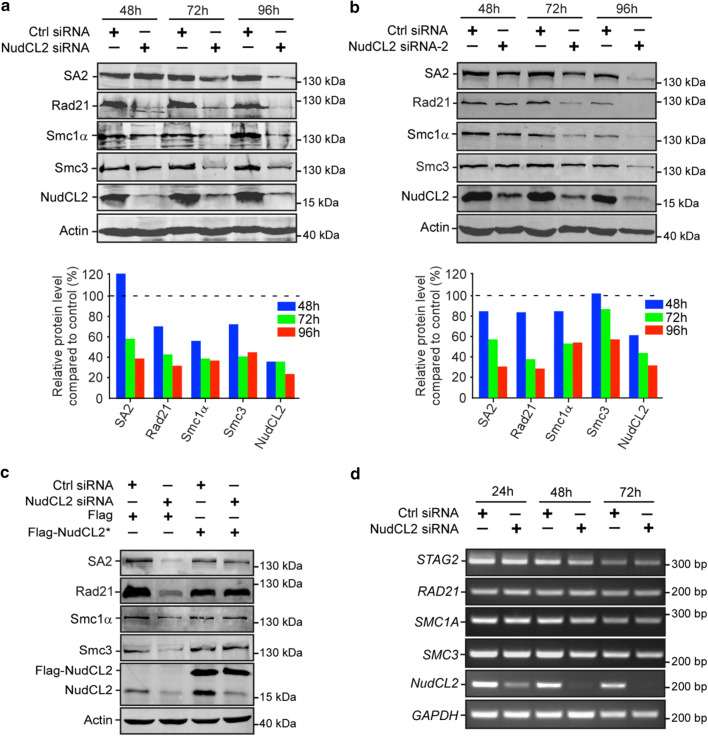
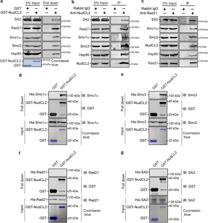
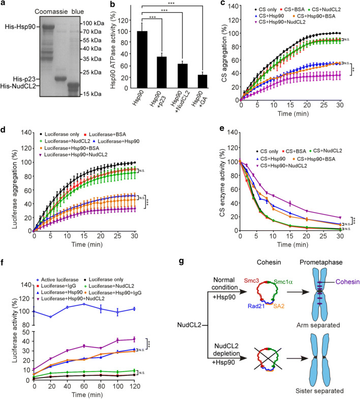
Sister chromatid cohesion plays a key role in ensuring precise chromosome segregation during mitosis, which is mediated by the multisubunit cohesin complex. However, the molecular regulation of cohesin subunits stability remains unclear. Here, we show that NudCL2 (NudC-like protein 2) is essential for the stability of cohesin subunits by regulating Hsp90 ATPase activity in mammalian cells. Depletion of NudCL2 induces mitotic defects and premature sister chromatid separation and destabilizes cohesin subunits that interact with NudCL2. Similar defects are also observed upon inhibition of Hsp90 ATPase activity. Interestingly, ectopic expression of Hsp90 efficiently rescues the protein instability and functional deficiency of cohesin induced by NudCL2 depletion, but not vice versa. Moreover, NudCL2 not only binds to Hsp90, but also significantly modulates Hsp90 ATPase activity and promotes the chaperone function of Hsp90. Taken together, these data suggest that NudCL2 is a previously undescribed Hsp90 cochaperone to modulate sister chromatid cohesion by stabilizing cohesin subunits, providing a hitherto unrecognized mechanism that is crucial for faithful chromosome segregation during mitosis.

Keywords: Cochaperone; Hsp90; NudCL2; Sister chromatid cohesion; cohesin.


Conflict of interest statement

The authors declare that they have no conflict of interest.

Figures

Fig. 1
Fig. 1
Depletion of NudCL2 causes mitotic defects. HeLa cells were transfected with either control or NudCL2 siRNA for 72 h and subjected to the following analyses: a Western blot analysis showed efficient suppression of NudCL2. Actin was used as a loading control. bd Immunofluorescence analysis displayed misalignment of chromosomes in NudCL2-depleted cells. Cells were stained with an anti-α-tubulin antibody and human CREST serum (b). The mitotic index was calculated (c). The percentage of mitotic cells with misaligned chromosomes was significantly higher in NudCL2-depleted cells than in control cells (d). eg Immunostaining revealed that NudCL2-depleted cells exhibiting misaligned chromosomes were positive for cyclin B1 staining. Cells were probed with an anti-cyclin B1 antibody and human CREST serum (e). Mitotic cells positive for cyclin B1 were calculated (f). The frequency of cyclin B1-positive mitotic cells with misaligned chromosomes was plotted (g). hm The spindle checkpoint proteins Bub1 and Mad2 were present at the kinetochores of NudCL2-depleted cells with misaligned chromosomes. Cells were stained with the indicated antibodies and human CREST serum. Mitotic cells positive for Bub1 (i) or Mad2 (l) were calculated. Mitotic cells with misaligned chromosomes with Bub1 (j) or Mad2 (m) signals were also counted. n Live imaging of HeLa cells stably expressing GFP-H2B showed that some chromosomes failed to congress to the metaphase plate in NudCL2-depleted cells. Time is shown as hh:mm. DNA was visualized with DAPI. Scale bars, 10 μm. Quantitative data are expressed as the mean ± SD (at least three independent experiments). More than 180 cells were counted in each experiment. ***p < 0.001, Student’s t test
Fig. 2
Fig. 2
NudCL2 depletion results in premature sister chromatid separation. HeLa cells transfected with the indicated siRNAs and vectors for 72 h were processed for the following analyses: a immunofluorescence analysis showed that NudCL2-depleted cells exhibited misaligned chromosomes with one CREST dot associated with only one green Hec1 dot at kinetochores. Cells were stained with an anti-Hec1 antibody and human CREST serum. DNA was visualized by DAPI. bf Chromosome spread assays with Giemsa staining revealed precocious sister chromatid separation in cells depleted of NudCL2. Western blot analysis displayed efficient suppression of NudCL2, Rad21 and Plk1 (b). Actin was used as a loading control. The cells were treated with colcemid for 2.5 h and subjected to chromosome spread analysis (c, d). Ectopic expression of RNAi-resistant NudCL2 (NudCL2*) effectively reversed the defects in sister chromatid separation induced by NudCL2 depletion (e, f). g, h Chromosome spread assay followed by immunofluorescence analysis with the indicated antibodies confirmed the precocious separation of sister chromatids in NudCL2-depleted cells. The cells grown on coverslips were treated with colcemid and subjected to chromosome spread and immunofluorescence analysis. DNA was stained with DAPI. Mitotic cells in chromosome spread experiments were categorized into four groups according to their chromosomal morphology: (1) arm separated, where the arms of sister chromatids were separated but connected at centromeres; (2) sister separated (separated sister chromatids), where sister chromatids were separated along the whole chromosome, but their pairing was maintained; (3) single chromosomes, where sister chromatids were completely separated and scattered; (4) arm closed, where sister chromatids were connected at both centromeres and arms. Mitotic cells with different chromosomal morphologies were calculated. Quantitative data are presented as the mean ± SD (at least three independent experiments). More than 150 cells were measured in each experiment. Bars, 10 μm. Higher magnifications of the boxed regions are displayed
Fig. 3
Fig. 3
Knockdown of NudCL2 leads to the degradation of cohesin subunits. HeLa cells transfected with the indicated siRNAs and plasmids were subjected to the following analyses: a, b Western blot analysis with the indicated antibodies revealed that NudCL2 depletion by two different NudCL2 siRNAs effectively decreased the protein levels of cohesin subunits. Actin, a loading control. Relative protein levels compared to the control at the same time point of post-transfection were measured using Image J software and shown at the bottom of each figure. c RNAi rescue experiments showed that ectopic expression of NudCL2 rescued the decreased protein levels of cohesin subunits. d Semiquantitative RT-PCR demonstrated that knockdown of NudCL2 had no obvious effect on the mRNA levels of cohesin subunits
Fig. 4
Fig. 4
Inhibition of Hsp90 induces defects in sister chromatid cohesion. HeLa cells treated with geldanamycin (GA) or DMSO for 48 h were used for the following analyses: a Western analysis with the indicated antibodies revealed that inhibition of Hsp90 by GA treatment obviously reduced the protein levels of cohesin subunits. Actin, a loading control. Relative protein levels compared to the control at the same time point of GA treatment were measured using Image J software and shown at the bottom. bd Immunofluorescence showed the misalignment of chromosomes in cells treated with GA. The cells were stained with an anti-α-tubulin antibody and CREST serum (b). The mitotic index was calculated (c). The percentage of mitotic cells with misaligned chromosomes was plotted (d). eg Immunostaining displayed that the majority of GA-treated cells with misaligned chromosomes were positive for cyclin B1. Cells were probed with the indicated antibodies (e). Cyclin B1-positive mitotic cells were quantified (f). The frequency of cyclin B1-positive mitotic cells with misaligned chromosomes was plotted (g). h Live imaging of HeLa cells stably expressing GFP-H2B showed that chromosomes failed to congress to the metaphase plate in GA-treated cells. DNA was visualized with DAPI. Bars, 10 μm. i, j Cells treated with GA exhibited premature sister chromatid separation. HeLa cells were subjected to chromosome spreads followed by Giemsa staining after treatment with colcemid for 2.5 h (i). Insets, high magnifications of the boxed areas. Mitotic cells with different chromosomal morphologies were counted by the method described in Fig. 2 (j). Quantitative data are expressed as the mean ± SD (at least three independent experiments). More than 180 cells were scored in each experiment. ***p < 0.001, Student’s t test
Fig. 5
Fig. 5
Ectopic expression of Hsp90 rescues the defects caused by NudCL2 depletion but not vice versa. HeLa cells transfected with the indicated siRNAs and vectors were treated with geldanamycin and subjected to Western blotting and chromosome spreads followed by Giemsa staining. Actin was used as a loading control. The percentages of mitotic cells with different chromosomal morphologies were measured by the method described in Fig. 2. Quantitative data are shown as the mean ± SD (at least three independent experiments). More than 170 cells were calculated in each experiment. a, b Exogenous expression of Hsp90 reversed the degradation of cohesin subunits and premature sister chromatid segregation in NudCL2-depleted cells. c, d Ectopic expression of NudCL2 was unable to rescue the decreased levels of cohesin subunits and defects in sister chromatid cohesion in cells with Hsp90 inhibition. e, f Depletion of NudCL2 had no synergistic effects with Hsp90 inhibition on cohesin subunit stability and sister chromatid cohesion
Fig. 6
Fig. 6
NudCL2 interacts with cohesin subunits and Hsp90 in vitro and in vivo. a GST pull-down assays showed that NudCL2 was associated with cohesin subunits and Hsp90 in vitro. Purified GST or GST-NudCL2 protein was incubated with HeLa cell lysates and subjected to immunoblotting. The inputs of GST and GST-NudCL2 were stained with Coomassie brilliant blue. b, c Endogenous NudCL2 bound to four cohesin subunits and Hsp90 in vivo. Total lysates of HeLa cells were immunoprecipitated with the indicated antibodies or IgGs and processed for Western blotting. dg NudCL2 protein directly interacted with Smc1α and Smc3 but not Rad21 or SA2 in vitro. His-tagged Rad21, SA2, Smc1α and Smc3 were expressed in Sf9 insect cells and purified with nickel-nitrilotriacetic acid beads. GST or GST-NudCL2 protein was incubated with each of the purified cohesin subunits and subjected to Western analysis with the indicated antibodies
Fig. 7
Fig. 7
NudCL2 enhances the chaperone activity of Hsp90. a Purified His-tagged p23, Hsp90, and NudCL2 proteins were subjected to SDS-PAGE with Coomassie blue staining. b NudCL2 inhibited the in vitro ATPase activity of Hsp90. Hsp90 ATPase activity was measured in the presence of p23, NudCL2 or geldanamycin and presented as the activity relative to that of only Hsp90. c, d NudCL2 suppressed the heat-induced aggregation of citrate synthase (CS) and luciferase with Hsp90. CS (c) and luciferase (d) were incubated with the indicated proteins for different times. Aggregation was determined by light scattering (370 nm) at each time point and calculated as the percentage of CS or luciferase aggregation after a 30 min incubation. e NudCL2 reduced the rate of heat-induced CS inactivation with Hsp90. CS was incubated with the indicated proteins at 43 °C for different times. The enzymatic activity of CS was detected at 30 °C by light scattering at 412 nm. All activities were expressed as a percentage of the initial activity of CS (0 min). f NudCL2 facilitated the refolding of luciferase after heat-induced aggregation. Luciferase was incubated at 42 °C for 15 min in the presence of the indicated proteins. The enzymatic activity of luciferase was monitored by a luminometer at each time point. The activity was calculated as a percentage of the activity of luciferase at 22 °C after a 15 min incubation. Quantitative data are presented as the mean ± SD (at least three independent experiments). ***p < 0.001, Student’s t test. g Working model for the role of NudCL2 in sister chromatid cohesion. NudCL2 stabilizes cohesin subunits by acting as an Hsp90 cochaperone to enhance cohesin stability. Depletion of NudCL2 induces cohesin subunits degradation and results in premature sister chromatid segregation during mitosis

References

    1. Nasmyth K, Haering CH. Cohesin: its roles and mechanisms. Annu Rev Genet. 2009;43:525–558. doi: 10.1146/annurev-genet-102108-134233. - DOI - PubMed
    1. Brooker AS, Berkowitz KM. The roles of cohesins in mitosis, meiosis, and human health and disease. Methods Mol Biol. 2014;1170:229–266. doi: 10.1007/978-1-4939-0888-2_11. - DOI - PMC - PubMed
    1. Watrin E, Schleiffer A, Tanaka K, Eisenhaber F, Nasmyth K, Peters JM. Human Scc4 is required for cohesin binding to chromatin, sister-chromatid cohesion, and mitotic progression. Curr Biol. 2006;16(9):863–874. doi: 10.1016/j.cub.2006.03.049. - DOI - PubMed
    1. Kueng S, Hegemann B, Peters BH, Lipp JJ, Schleiffer A, Mechtler K, Peters JM. Wapl controls the dynamic association of cohesin with chromatin. Cell. 2006;127(5):955–967. doi: 10.1016/j.cell.2006.09.040. - DOI - PubMed
    1. Zhang J, Shi X, Li Y, Kim BJ, Jia J, Huang Z, Yang T, Fu X, Jung SY, Wang Y, Zhang P, Kim ST, Pan X, Qin J. Acetylation of Smc3 by Eco1 is required for S phase sister chromatid cohesion in both human and yeast. Mol Cell. 2008;31(1):143–151. doi: 10.1016/j.molcel.2008.06.006. - DOI - PubMed

MeSH terms

LinkOut - more resources