

New generation of bioreactors that advance extracellular matrix modelling and tissue engineering
- PMID: 30368691
- PMCID: PMC6313369
- DOI: 10.1007/s10529-018-2611-7
New generation of bioreactors that advance extracellular matrix modelling and tissue engineering
Abstract
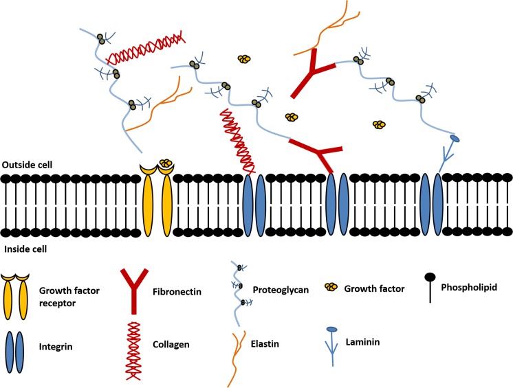
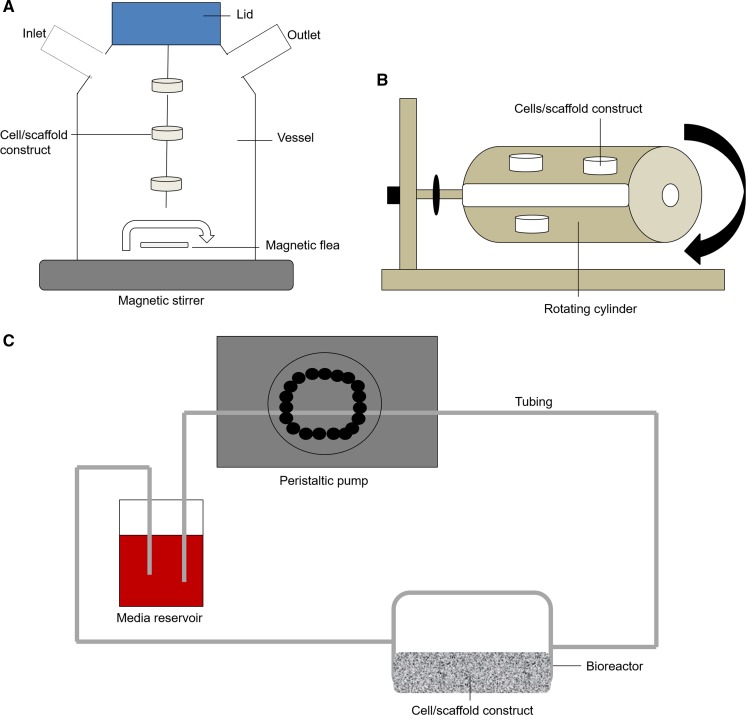
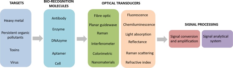
Bioreactors hold a lot of promise for tissue engineering and regenerative medicine applications. They have multiple uses including cell cultivation for therapeutic production and for in vitro organ modelling to provide a more physiologically relevant environment for cultures compared to conventional static conditions. Bioreactors are often used in combination with scaffolds as the nutrient flow can enhance oxygen and diffusion throughout the 3D constructs to prevent the formation of necrotic cores. A variety of scaffolds have been fabricated to achieve a structural architecture that mimic native extracellular matrix. Future developments of in vitro models will incorporate the ability to non-invasively monitor the cellular microenvironment to enhance the understanding of in vitro conditions. This review details current advancements in bioreactor and scaffold systems and provides insight on how in vitro models can be augmented for future biomedical applications.
Keywords: Bio-sensing; Bioreactor; Extracellular matrix; Nanosensors; Regenerative medicine; Scaffolds; Tissue engineering.
Conflict of interest statement
The authors declare that they have no conflict of interest.
Figures


Similar articles
-
Engineered channels enhance cellular density in perfused scaffolds.Acta Biomater. 2011 Nov;7(11):3896-904. doi: 10.1016/j.actbio.2011.06.037. Epub 2011 Jun 28. Acta Biomater. 2011. PMID: 21745609
-
A Perfusion Bioreactor System for Cell Seeding and Oxygen-Controlled Cultivation of Three-Dimensional Cell Cultures.Tissue Eng Part C Methods. 2018 Oct;24(10):585-595. doi: 10.1089/ten.TEC.2018.0204. Tissue Eng Part C Methods. 2018. PMID: 30234443 Free PMC article.
-
Modulation of cell differentiation in bone tissue engineering constructs cultured in a bioreactor.Adv Exp Med Biol. 2006;585:225-41. doi: 10.1007/978-0-387-34133-0_16. Adv Exp Med Biol. 2006. PMID: 17120788 Review.
-
Concentric cylinder bioreactor for production of tissue engineered cartilage: effect of seeding density and hydrodynamic loading on construct development.Biotechnol Prog. 2003 Mar-Apr;19(2):510-21. doi: 10.1021/bp0256519. Biotechnol Prog. 2003. PMID: 12675595
-
Review: bioreactor design towards generation of relevant engineered tissues: focus on clinical translation.J Tissue Eng Regen Med. 2018 Jan;12(1):e7-e22. doi: 10.1002/term.2270. Epub 2017 Apr 3. J Tissue Eng Regen Med. 2018. PMID: 28374578 Review.
Cited by
-
Cell-scaffold interactions in tissue engineering for oral and craniofacial reconstruction.Bioact Mater. 2022 Nov 8;23:16-44. doi: 10.1016/j.bioactmat.2022.10.029. eCollection 2023 May. Bioact Mater. 2022. PMID: 36406245 Free PMC article.
-
Decellularization in Tissue Engineering and Regenerative Medicine: Evaluation, Modification, and Application Methods.Front Bioeng Biotechnol. 2022 Apr 25;10:805299. doi: 10.3389/fbioe.2022.805299. eCollection 2022. Front Bioeng Biotechnol. 2022. PMID: 35547166 Free PMC article. Review.
-
Application of decellularization methods for scaffold production: advantages, disadvantages, biosafety and modifications.Front Bioeng Biotechnol. 2025 Jun 18;13:1621641. doi: 10.3389/fbioe.2025.1621641. eCollection 2025. Front Bioeng Biotechnol. 2025. PMID: 40606910 Free PMC article. Review.
-
Optimization of Topography and Surface Properties of Polyacrylonitrile-Based Electrospun Scaffolds via Nonoclay Concentrations and its Effect on Osteogenic Differentiation of Human Mesenchymal Stem Cells.Iran J Pharm Res. 2021 Fall;20(4):385-504. doi: 10.22037/ijpr.2021.115119.15208. Iran J Pharm Res. 2021. PMID: 35194454 Free PMC article.
-
3D Cell Cultures: Evolution of an Ancient Tool for New Applications.Front Physiol. 2022 Jul 22;13:836480. doi: 10.3389/fphys.2022.836480. eCollection 2022. Front Physiol. 2022. PMID: 35936888 Free PMC article. Review.
References
-
- Acosta MA, Kostov Y, Leach JB (2007) Microfluidic bioreactors as tools for monitoring cell microenvironment. 200–200
-
- Akbarzadeh R, Yousefi AM. Effects of processing parameters in thermally induced phase separation technique on porous architecture of scaffolds for bone tissue engineering. J Biomed Mater Res. 2014;102(6):1304–1315. - PubMed
-
- Askari P, Zahedi P, Rezaeian I. Three-layered electrospun PVA/PCL/PVA nanofibrous mats containing tetracycline hydrochloride and phenytoin sodium: a case study on sustained control release, antibacterial, and cell culture properties. J Appl Polym Sci. 2016;133:1–9.
-
- Attia M, Santerre JP, Kandel RA. The response of annulus fibrosus cell to fibronectin-coated nanofibrous polyurethane-anionic dihydroxyoligomer scaffolds. Biomaterials. 2011;32:450–460. - PubMed
Publication types
MeSH terms
Grants and funding
LinkOut - more resources
Full Text Sources
Other Literature Sources
