. 2018 Dec;233(6):828-842.
doi: 10.1111/joa.12883. Epub 2018 Oct 14.

The missing segment of the autopod 1st ray: new insights from a morphometric study of the human hand

Affiliations

The missing segment of the autopod 1st ray: new insights from a morphometric study of the human hand

Ugo E Pazzaglia et al. J Anat. 2018 Dec.

Abstract

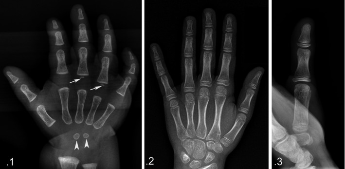
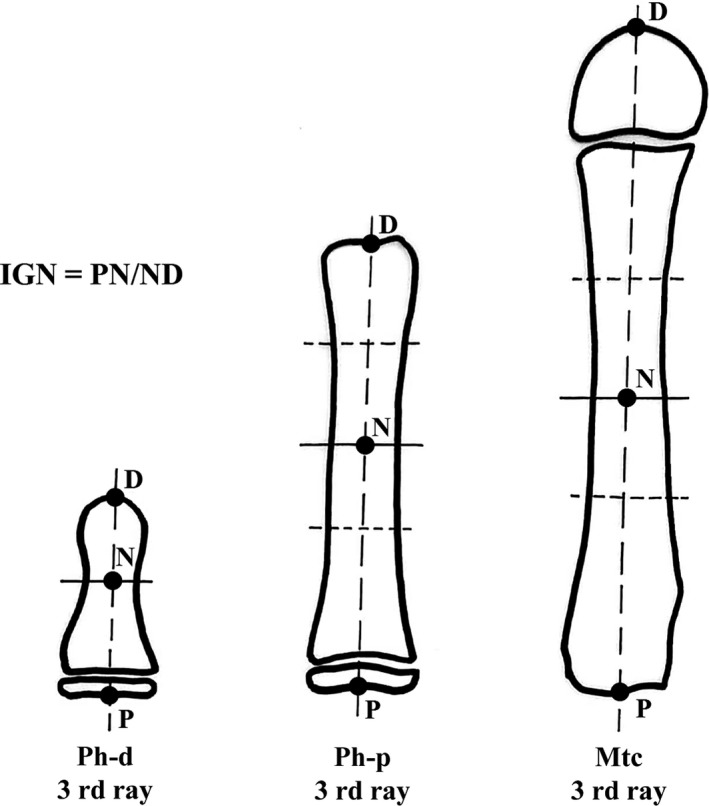
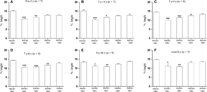
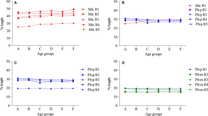
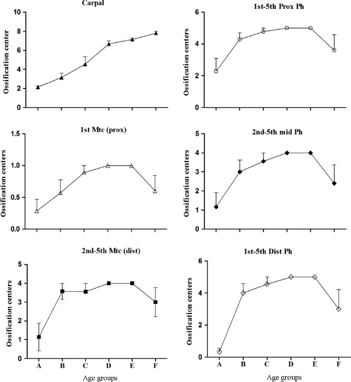
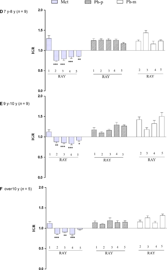
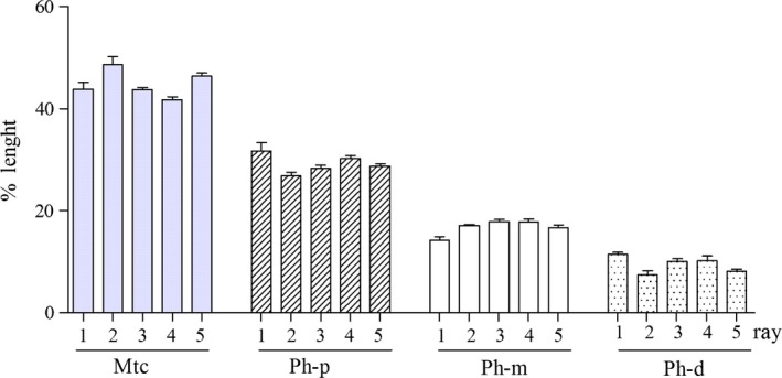
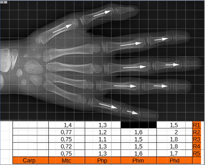
Whether the 1st segment of the human autopod 1st ray is a 'true' metapodial with loss of the proximal or mid phalanx or the original basal phalanx with loss of the metacarpal has been a long-lasting discussion. The actual knowledge of the developmental pattern of upper autopod segments at a fetal age of 20-22 weeks, combined with X-ray morphometry of normal long bones of the hand in the growing ages, was used for analysis of the parameters, percentage length, position of epiphyseal ossification centers and proximal/distal growth rate. The symmetric growth pattern in the fetal anlagen changed to unidirectional in the postnatal development in relation to epiphyseal ossification formation. The percentage length assessment, the distribution of the epiphyseal ossification centers, and differential proximal/distal growth rate among the growing hand segments supported homology of most proximal segment of the thumb with the 2nd-5th proximal phalanges and that of the proximal phalanx of the thumb with the 2nd-5th mid phalanges in the same hand. Published case reports of either metanalysis of 'triphalangeal thumb' and 'proximal/distal epiphyseal ossification centers' were used to support the applied morphometric methodology; in particular, the latter did not give evidence of growth pattern inversion of the proximal segment of the thumb. The presented data support the hypothesis that during evolution, the lost segment of the autopod 1st ray is the metacarpal.

Keywords: autopod fetal anlage growth; fetal ossification pattern; morphometric and patterning homology; postnatal ossification pattern.


Figures

Figure 1
Figure 1
(1) Right‐hand X‐ray, a‐p projection (age 8 months, group A). Early stage of ossification with two centers of the carpal short bone anlagen and with basal, epiphyseal ossification centers of the 3rd and 4th ray proximal phalanges. The thumb bone segments are taken in an oblique projection, which is not comparable for shape analysis with those of the 2nd and 5th rays. (2) Right‐hand X‐ray, a‐p projection (age 13 years, group F). Advanced stage of ossification with all eight carpal bone ossification centers and the presence of all the long bone ossification centers: proximal position of the 1st–5th phalanges and inverted position of the thumb metacarpal to the 2nd–5th metacarpals. The shape of the thumb ossification center can be classified as flattened even if it is thicker than the phalangeal center, but it certainly is not similar to the round‐shaped distal epiphyses of the 2nd–5th metacarpals. The thumb bone segments are taken in oblique projection as in age group A. (3) Hand X‐ray, 1st ray a‐p projection (age 9 years, group E). Shape analysis of the thumb segments in this projection allows comparison with the other ray segments.
Figure 2
Figure 2
Graphic illustration of the IGR) measurement method in postnatal long bones (see details in Materials and methods). This assessment was applicable only in segments with a well‐developed epiphyseal ossification center (age groups D–F).
Figure 3
Figure 3
The 1st ray distal phalanx mean percentage length (measured on the total length of the 3rd ray) was compared with the mean percentage length of the 2nd–5th ray distal phalanges (measured on the total length of each ray). The result was significantly higher than that for the 2nd–3rd ray distal phalanges in all age groups A–F but not significantly different than that of the 4th–5th rays of age groups C–D. The typology of the 1st ray distal phalanx cannot be questioned because of the characterizing apical tuft morphology. Therefore, the observed differences documented a ‘true’ major growth of the latter segment vs. the 2nd–3rd rays; this is independent of the percentage measurement method, which assumed that the reference to the total length of the 3rd ray corrected the bias due to the missing segment of the thumb (* P < 0.05; ** P < 0.01; *** P < 0.001).
Figure 4
Figure 4
1 (A,B) Graphic profile of ray 1–5 total length of the metacarpal percentage length (ray 1 measured on ray 3 total length, rays 2–5 on the total length of each ray) in age groups A–F. This documents the percentage length dishomology of Mtc R1 (red) with respect to Mtcs R2‐R5 (red) and the homology of the same Mtc R1 (red) with respect to the percentage length of Ph‐p R2‐R5 (blue). (C,D) Corresponding graphic profile of R1‐R5 metacarpal percentage length (R1 measured on R3 total length, R2–R5 on the total length of each ray) documenting the percentage length dishomology of Ph‐p R1 (blue) with respect to Ph‐p R2‐R5 (blue) and the homology of the same Ph‐p R1 (green) with respect to percentage length of Ph‐m R2‐R5 (blue).
Figure 5
Figure 5
The regular progression of the number of carpal ossification centers with age confirmed the current use in the clinical assessment of skeletal age (Vogt & Vickers, 1938; Greunlich & Pyle, 1959). The different slope of the tubular bone epiphyseal ossification center number among the age groups is representative of variability of the time of appearance in epiphyseal center ossification. The reduction in number between age groups A and F corresponds to fusion with the ossified diaphyses.
Figure 6
Figure 6
Proximal‐distal IGR) compared among R1–R5 metacarpals (Mtc), proximal phalanges (Ph‐p) and mid phalanges (Ph‐m) in age groups D–E. This parameter was not assessable in age groups A–C. With reference to IGR ≅ 1 corresponding to symmetric, bidirectional growth, the index was inverted at the passage from the 1st and the 2nd metacarpals with an evident relationship with the epiphyseal ossification center position (and later growth plate cartilage). Significant differences in proximal and mid phalanges (not reported in the histograms) but without inversion. *P < 0.05; **P < 0.01; *** P < 0.001 vs. R1 Mtc.
Figure 7
Figure 7
Triphalangeal thumb metanalysis. Comparison of the mean percentage length (measured on the total length of each ray) of 1st–5th ray metacarpals (Mtc), proximal (Ph‐p), mid (Ph‐m) and distal (Ph‐d) phalanges of TPT series (mean ± SEM of eight subjects). There was no significant difference when each segment type is considered in the transverse sequence R1–R5. The percentage length in all rays decreases from metacarpal to distal phalanges.
Figure 8
Figure 8
Image of triphalangeal thumb of the right and left hand of the mother (.1) and her newborn (.2) reported by Heiss (1953) and reproduced from Zeitschrift fur Anatomie und Entwicklungsgeschicte with permission of Springer Nature (licence no. 4334811065195).
Figure 9
Figure 9
(top) Image of the hand with the widest distribution of proximal and distal epiphyseal ossification centers, reported in two siblings by de Jong et al. (2014) and reproduced from The Journal of Hand Surgery with permission of Elsevier (licence no. 4280070488758). (bottom) Table reporting the IGR calculation of each hand segment.

References

    1. Almecija S, Smaers IB, Jungers WL (2015) The evolution of human ape hand proportion. Nat Commun. 6, 8717 10.1038/ncomms8717. - DOI - PMC - PubMed
    1. Bever GS, Gauthier JA, Wagner GP (2011) Finding the frame shift, digit loss developmental variability, and the origin of the avian hand. Evol Dev 13, 269–279. - PubMed
    1. Bland JM, Altman DG (2010) Statistical methods for assessing agreement between two methods of clinical measurement. Int J Nurs Stud 47, 931–936. - PubMed
    1. Blauth W (1967) Der hypoplastischeDaumen. Arch OrthopUnfallchir 62, 225–246. - PubMed
    1. Brailsford JF (1943) Variations in the ossifications of the one of the hand. J Anat 77, 170–175. - PMC - PubMed

Publication types