

Axonal swellings and spheroids: a new insight into the pathology of neurocysticercosis
- PMID: 30368965
- PMCID: PMC6482075
- DOI: 10.1111/bpa.12669
Axonal swellings and spheroids: a new insight into the pathology of neurocysticercosis
Abstract
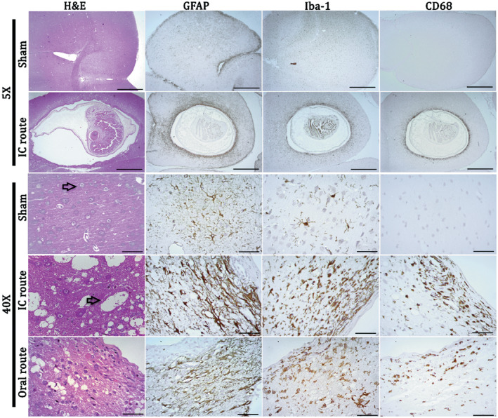
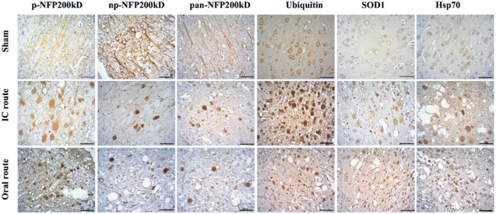
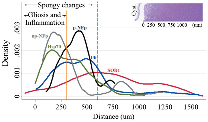
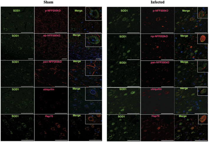
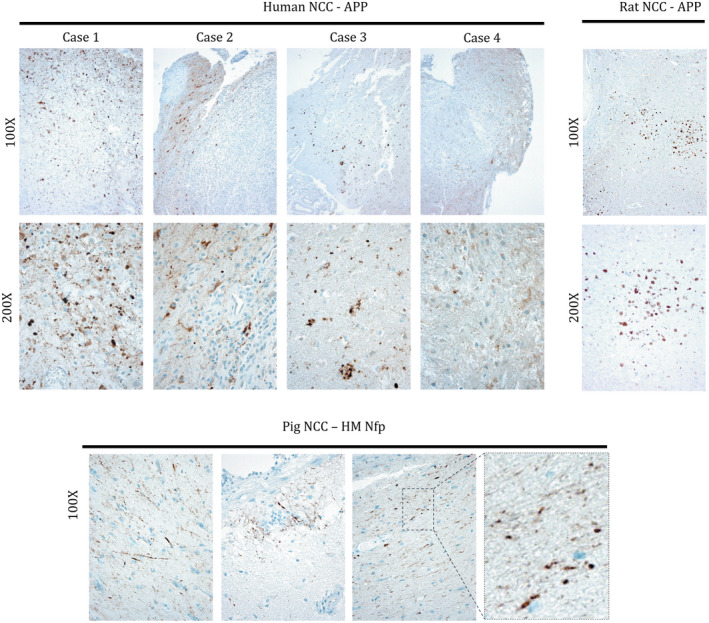
Neurocysticercosis is a parasitic brain disease caused by the larval form (Cysticercus cellulosae) of Taenia solium and is the leading cause of preventable epilepsy worldwide. However, the pathophysiology and relation to the wide range of clinical features remains poorly understood. Axonal swelling is emerging as an important early pathological finding in multiple neurodegenerative diseases and as a cause of brain injury, but has not been well described in neurocysticercosis. Histological analysis was performed on human, rat and porcine NCC brain specimens to identify axonal pathology. Rat infection was successfully carried out via two routes of inoculation: direct intracranial injection and oral feeding. Extensive axonal swellings, in the form of spheroids, were observed in both humans and rats and to a lesser extent in pigs with NCC. Spheroids demonstrated increased immunoreactivity to amyloid precursor protein and neurofilament indicating probable impairment of axonal transport. These novel findings demonstrate that spheroids are present in NCC which is conserved across species. Not only is this an important contribution toward understanding the pathogenesis of NCC, but it also provides a model to analyze the association of spheroids with specific clinical features and to investigate the reversibility of spheroid formation with antihelminthic treatment.
Keywords: APP; T. solium oncospheres; neurocysticercosis; neurofilament; spheroids.
© 2018 International Society of Neuropathology.
Conflict of interest statement
The authors declare that they have no conflict of interest.
Figures
Comment in
-
Letter in response to Del Brutto, axonal swelling and spheroids in Taenia solium neurocysticercosis.Brain Pathol. 2019 May;29(3):319. doi: 10.1111/bpa.12700. Epub 2019 Feb 11. Brain Pathol. 2019. PMID: 30637848 Free PMC article. No abstract available.
-
Axonal swelling and spheroids in Taenia solium neurocysticercosis.Brain Pathol. 2019 May;29(3):320. doi: 10.1111/bpa.12701. Epub 2019 Feb 11. Brain Pathol. 2019. PMID: 30637855 Free PMC article. No abstract available.
References
-
- Alvarez JI, Colegial CH, Castaño CA, Trujillo J, Teale JM, Restrepo BI (2002) The human nervous tissue in proximity to granulomatous lesions induced by Taenia solium metacestodes displays an active response. J Neuroimmunol 127:139–144. - PubMed
-
- Arora N, Tripathi S, Kumar P, Mondal P, Mishra A, Prasad A (2017) Recent advancements and new perspectives in animal models for Neurocysticercosis immunopathogenesis. Parasite Immunol 39:e12439. - PubMed
-
- Born HA (2015) Seizures in Alzheimer’s disease. Neuroscience 286:251–263. - PubMed
Publication types
MeSH terms
Grants and funding
LinkOut - more resources
Full Text Sources
