. 2018 Dec;30(50):e1805557.
doi: 10.1002/adma.201805557. Epub 2018 Oct 11.

Nanoparticle-Laden Macrophages for Tumor-Tropic Drug Delivery

Affiliations

Nanoparticle-Laden Macrophages for Tumor-Tropic Drug Delivery

Weizhong Zhang et al. Adv Mater. 2018 Dec.

Erratum in

  • Nanoparticle-Laden Macrophages for Tumor-Tropic Drug Delivery.
    Zhang W, Wang M, Tang W, Wen R, Zhou S, Lee C, Wang H, Jiang W, Delahunty IM, Zhen Z, Chen H, Chapman M, Wu Z, Howerth EW, Cai H, Li Z, Xie J. Zhang W, et al. Adv Mater. 2022 Feb;34(6):e2109925. doi: 10.1002/adma.202109925. Adv Mater. 2022. PMID: 35141954 No abstract available.

Abstract

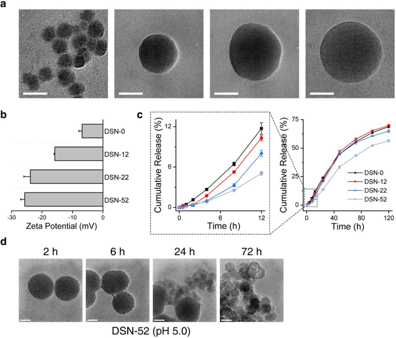
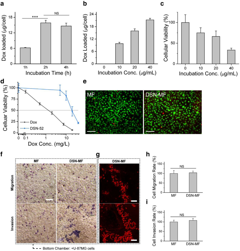
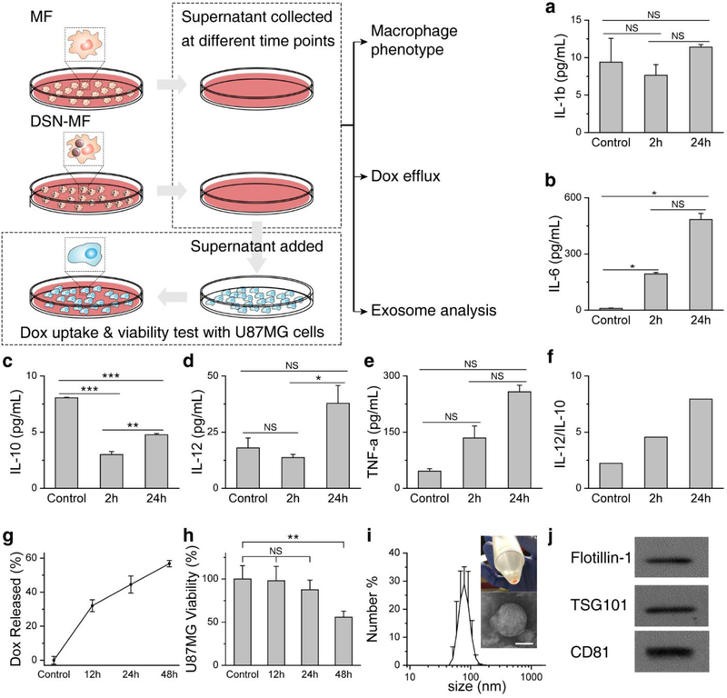
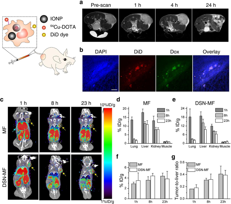
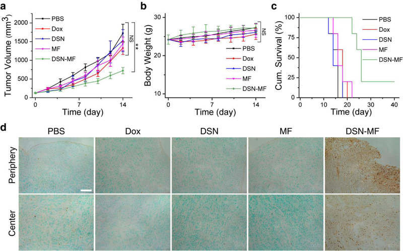
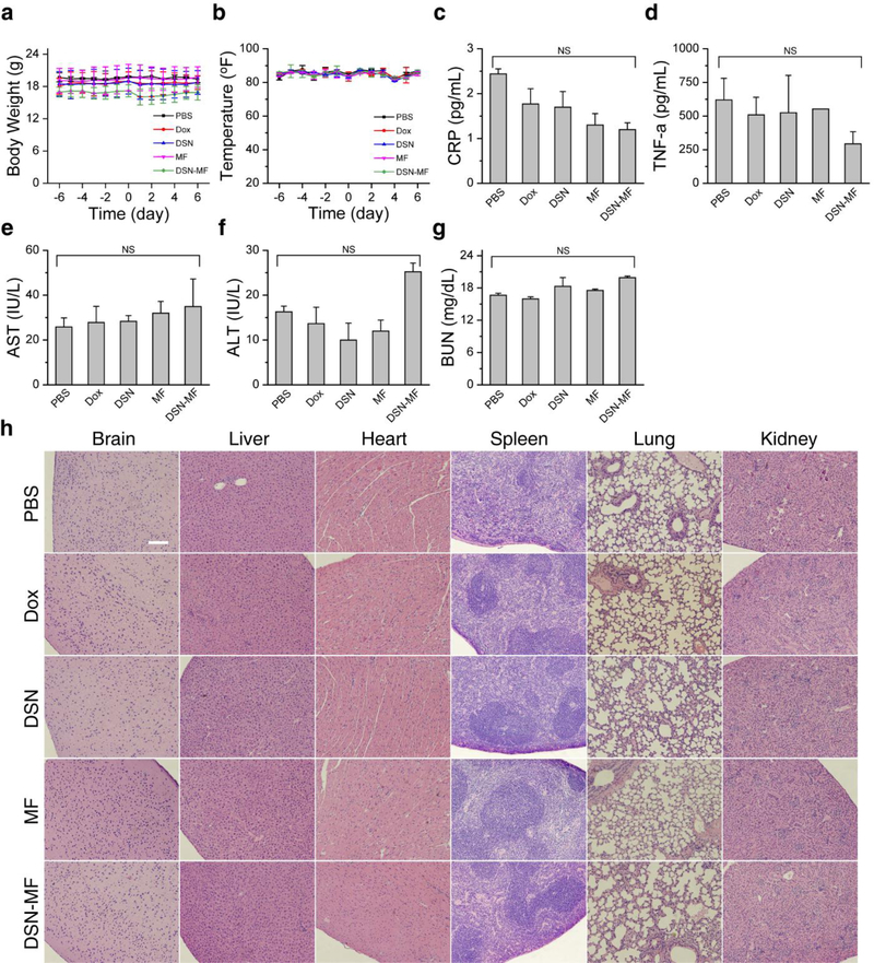
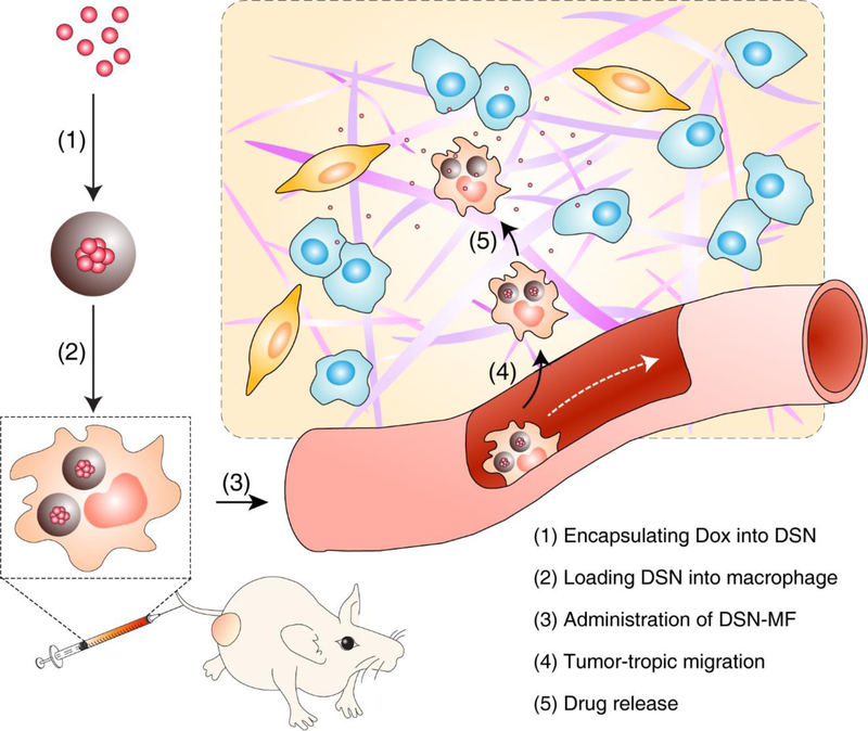
Macrophages hold great potential in cancer drug delivery because they can sense chemotactic cues and home to tumors with high efficiency. However, it remains a challenge to load large amounts of therapeutics into macrophages without compromising cell functions. This study reports a silica-based drug nanocapsule approach to solve this issue. The nanocapsule consists of a drug-silica complex filling and a solid silica sheath, and it is designed to minimally release drug molecules in the early hours of cell entry. While taken up by macrophages at high rates, the nanocapsules minimally affect cell migration in the first 6-12 h, buying time for macrophages to home to tumors and release drugs in situ. In particular, it is shown that doxorubicin (Dox) as a representative drug can be loaded into macrophages up to 16.6 pg per cell using this approach. When tested in a U87MG xenograft model, intravenously (i.v.) injected Dox-laden macrophages show comparable tumor accumulation as untreated macrophages. Therapy leads to efficient tumor growth suppression, while causing little systematic toxicity. This study suggests a new cell platform for selective drug delivery, which can be readily extended to the treatment of other types of diseases.

Keywords: cancer; cell-mediated drug delivery; doxorubicin; glioblastoma; macrophages; nanoparticles.


Figures

Figure 1.
Figure 1.
Physical characterizations of DSN nanoparticles. a) TEM images and b) zeta potential of DSN-0, DSN-12, DSN-22, and DSN-52 nanoparticles. c) Drug release profiles of DSN-0, DSN-12, DSN-22, and DSN-52 nanoparticles, measured at pH 5.0. d) TEM images of DSN-52 nanoparticles after incubating in a pH 5.0 solution for different times. Scale bars, 50 nm.
Figure 2.
Figure 2.
DSN-52 nanoparticles uptake by macrophages (RAW264.7 cells). a) Intracellular Dox contents, measured at 1, 2, and 4 hours’ incubation with DSN-52 nanoparticles. ***, p<0.001. NS, not significant. b) Intracellular Dox contents, measured when the initial DSN-52 Dox concentration was 0, 10, 20, and 40 μg mL−1. The incubation time was fixed at 2 h. c) Cell viability at 12 h via MTT assay. The cells were first incubated with DSN-52 at 0, 10, 20, and 40 μg mL−1 (Dox concentration) for 2 h. After PBS washing, fresh growth medium was added, and cell viability was measured at 12 h by MTT assay. d) Cell viability at 24 h via MTT assay. Dox (black curve), RAW264.7 cells were incubated with free Dox for 24 h. DSN-52 (blue curve), RAW264.7 cells were laden with DSN-52 and then incubated in normal growth medium for 24 h. e) Live and Dead cell assay results of DSN-MF and MF cells at 2 h. Green, living cells; red, dead cells. Scale bars, 100 μm. f) Transmigration assay. DSN-MF or MF cells were loaded onto the top of a transwell chamber, whilst U87MG cells were seeded at the bottom. Macrophages were stained into blue color via Giemsa staining. Scale bars, 100 μm. g) Fluorescence microscopic images of invaded/migrated DSN-MF cells, the experimental conditions were the same as those in f. Scale bars, 100 μm. Percentages of DSN-MF and MF cells that had h) migrated and i) invaded. NS, not significant.
Figure 3.
Figure 3.
Impact of DSN loading on macrophage phenotypes. Secretion of a) IL-1β, b) IL-6, c) IL-10, d) IL-12, and e) TNF-α from DSN-MF at 2 and 24 h. MF (untreated RAW264.7 cells) served as controls. f) IL-12/IL-10 ratio at 2 and 24 h. g) Percentage of Dox released from DSN-MF at different times (Dox retained in cell debris is excluded by centrifugation). h) Cell viability assay results with U87MG cells. Supernatants taken from DSN-MF culture dishes at different time points were added to a separate plate grown with U87MG cells. Cell viability was measured after 48 h incubation. *, P<0.05; **, P<0.01; ***, P<0.001; NS, not significant. i) Hydrodynamic size of exosomes via DLS analysis (z-average size = 97.35 nm, PDI = 0.127). Exosomes were collected from DSN-MF supernatant at 48 h via centrifugations. An inset photograph of the resulting exosomes and a negative-stained TEM image were also shown. Scale bar, 50 nm. j) Western blot analysis of exosome lysates. Flotilin-1, TSG101, and CD81, three markers of exosomes, were detected.
Figure 4.
Figure 4.
In vivo tumor targeting of DSN-MF, evaluated in nude mice bearing subcutaneously inoculated U87MG tumors. a) Axial T2 MR images, acquired at 0, 1, 4, and 24 h post i.v. injection of DSN-MF cells. The cells were pre-loaded with iron oxide nanoparticles. b) Confocal microscopic images of tumor cryo-sections using the z-stack scan mode (step = 2 μm). DSN-MF cells were pre-labeled with DiD. Red, DiD; green, Dox; blue, cell nuclei. Scale bars, 50 μm. c) Decay-corrected whole-body coronal PET images, acquired at 1, 8, and 23 h post injection. DSN-MF or MF cells were labeled with 64Cu-PTSM. Tumor area was highlighted with yellow cycles; lung area was highlighted using cyan cycle. d, e) Distribution of d) MF cells and e) DSN-MF cells in the lung, liver, kidney, and muscle at different time points. f) Tumor uptake of MF and DSN-MF cells at different times. g) Tumor-to-liver ratios of MF and DSN-MF cells, based on images results in c.
Figure 5.
Figure 5.
Therapy studies with U87MG tumor bearing mice. Animals were randomized to receive one dose i.v. injection of either PBS, free Dox (3 mg Dox kg−1), DSN-52 (3 mg Dox kg1), RAW264.7 cells (MF, ~4 × 10⁶ cells per mouse), or DSN-MF (3 mg Dox kg−1, ~4 × 10⁶ cells per mouse). a) Tumor growth curves. b) Body weight changes. c) Kaplan-Meier plot of animal survival. d) In situ Apoptosis staining (Abcam) analysis of cryo-sectioned tumor tissues at 24 h post treatments. Cytoplasm region was counterstained into green color by methyl green; nuclei of apoptotic cells were counterstained into dark brown dots by diaminobenzidine. Scale bar, 50 μm.
Figure 6.
Figure 6.
Toxicity studies. a) Animal body weight changes. b) Animal rectal temperature changes. There was a small degree of weight loss in Dox, DSN-52, and DSN-MF group, which was recovered within 5 days. Mice were euthanized on Day 7 for H&E and plasma protein marker analysis: c) Plasma CRP, d) TNF-α, e, f) AST, ALT levels, and g) BUN levels. For DSNMF, all the indices were in the normal range. h) H&E staining of major organs, which were collected on Day 7 post treatments. Except for a small degree of elevated leukocyte infiltration, no pathological changes were observed for the DSN-MF group. Scale bar, 100 μm.
Scheme 1.
Scheme 1.. Nanocapsule-laden macrophages for drug delivery to tumors.
(1) Antineoplastic drug, in this particular case Dox, was first loaded into a carefully tailored nanocapsule called drug-silica nanocomplex (DSN); (2) DSN nanoparticles were engulfed by macrophages ex vivo; (3) DSN-laden macrophages (DSN-MF) were i.v. injected to a tumor bearing mouse; (4) chemotactic migration of DSN-MF to tumors; (5) DSN-MF releases Dox inside tumor to selectively kill cancer cells.

References

    1. Wang Q, Cheng H, Peng H, Zhou H, Li PY, Langer R, Adv. Drug Deliv. Rev 2015, 91, 125. - PubMed
    1. Su Y, Xie Z, Kim GB, Dong C, Yang J, ACS Biomater. Sci. Eng 2015, 1, 201. - PMC - PubMed
    1. Anselmo AC, Mitragotri S, J. Control. Release 2014, 190, 531. - PMC - PubMed
    1. Yi L, Xiao H, Xu M, Ye X, Hu J, Li F, Li M, Luo C, Yu S, Bian X, Feng H, J. Neuroimmunol 2011, 232, 75. - PubMed
    1. Singh A, Talekar M, Raikar A, Amiji M, J. Control. Release 2014, 190, 515. - PubMed

MeSH terms

LinkOut - more resources