

Integrated optical coherence tomography and multielement ultrasound transducer probe for shear wave elasticity imaging of moving tissues
- PMID: 30369107
- PMCID: PMC6210783
- DOI: 10.1117/1.JBO.23.10.105006
Integrated optical coherence tomography and multielement ultrasound transducer probe for shear wave elasticity imaging of moving tissues
Abstract
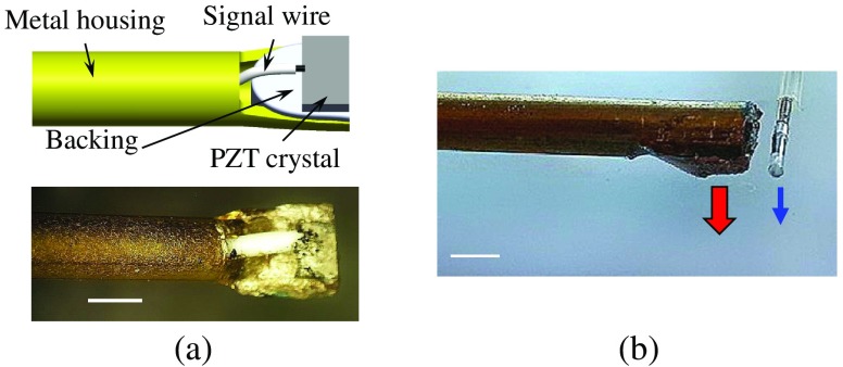
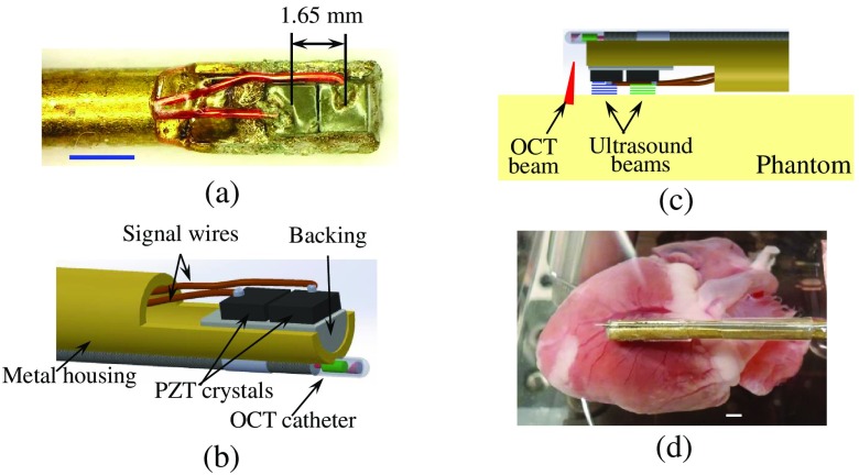
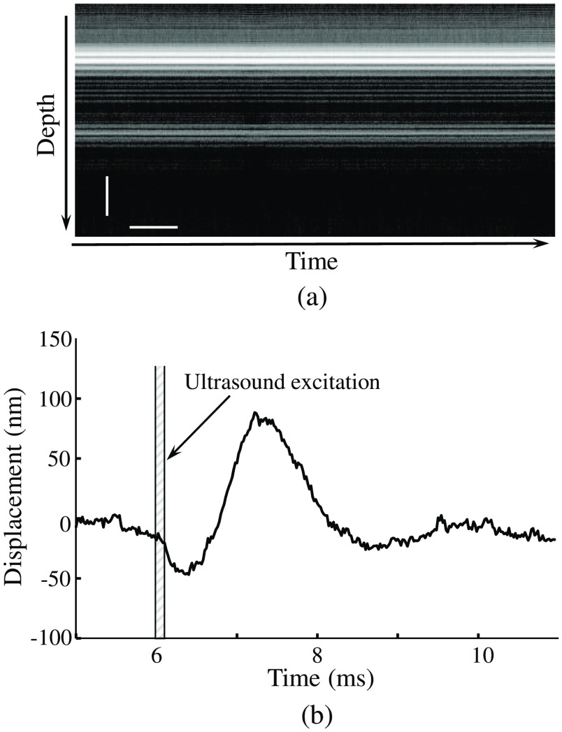
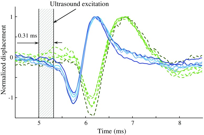
Accurate measurements of microelastic properties of soft tissues in-vivo using optical coherence elastography can be affected by motion artifacts caused by cardiac and respiratory cycles. This problem can be overcome using a multielement ultrasound transducer probe where each ultrasound transducer is capable of generating acoustic radiation force (ARF) and, therefore, creating shear waves in tissue. These shear waves, produced during the phase of cardiac and respiratory cycles when tissues are effectively stationary, are detected at the same observation point using phase-sensitive optical coherence tomography (psOCT). Given the known distance between the ultrasound transducers, the speed of shear wave propagation can be calculated by measuring the difference between arrival times of shear waves. The combined multitransducer ARF/psOCT probe has been designed and tested in phantoms and ex-vivo studies using fresh rabbit heart. The measured values of shear moduli are in good agreement with those reported in literature. Our results suggest that the developed multitransducer ARF/psOCT probe can be useful for many in-vivo applications, including quantifying the microelasticity of cardiac muscle.
Keywords: acoustic radiation force; cardiac muscle; multielement ultrasound transducer probe; optical coherence tomography and elastography; shear wave elasticity imaging.
(2018) COPYRIGHT Society of Photo-Optical Instrumentation Engineers (SPIE).
Figures

Similar articles
-
From supersonic shear wave imaging to full-field optical coherence shear wave elastography.J Biomed Opt. 2013 Dec;18(12):121514. doi: 10.1117/1.JBO.18.12.121514. J Biomed Opt. 2013. PMID: 24357549
-
Quantitative shear-wave optical coherence elastography with a programmable phased array ultrasound as the wave source.Opt Lett. 2015 Nov 1;40(21):5007-10. doi: 10.1364/OL.40.005007. Opt Lett. 2015. PMID: 26512505
-
Probe Oscillation Shear Elastography (PROSE): A High Frame-Rate Method for Two-Dimensional Ultrasound Shear Wave Elastography.IEEE Trans Med Imaging. 2016 Sep;35(9):2098-106. doi: 10.1109/TMI.2016.2550007. Epub 2016 Apr 4. IEEE Trans Med Imaging. 2016. PMID: 27076352 Free PMC article.
-
Visualizing ultrasonically induced shear wave propagation using phase-sensitive optical coherence tomography for dynamic elastography.Opt Lett. 2014 Feb 15;39(4):838-41. doi: 10.1364/OL.39.000838. Opt Lett. 2014. PMID: 24562220 Free PMC article.
-
Shear modulus imaging by direct visualization of propagating shear waves with phase-sensitive optical coherence tomography.J Biomed Opt. 2013 Dec;18(12):121509. doi: 10.1117/1.JBO.18.12.121509. J Biomed Opt. 2013. PMID: 24213539 Free PMC article.
Cited by
-
In vivo endoscopic optical coherence elastography based on a miniature probe.Biomed Opt Express. 2024 Jun 12;15(7):4237-4252. doi: 10.1364/BOE.521154. eCollection 2024 Jul 1. Biomed Opt Express. 2024. PMID: 39022537 Free PMC article.
-
Four-dimensional (4D) phase velocity optical coherence elastography in heterogeneous materials and biological tissue.Biomed Opt Express. 2020 Jun 18;11(7):3795-3817. doi: 10.1364/BOE.394835. eCollection 2020 Jul 1. Biomed Opt Express. 2020. PMID: 33014567 Free PMC article.
-
Introduction to optical coherence elastography: tutorial.J Opt Soc Am A Opt Image Sci Vis. 2022 Mar 1;39(3):418-430. doi: 10.1364/JOSAA.444808. J Opt Soc Am A Opt Image Sci Vis. 2022. PMID: 35297425 Free PMC article.
-
Assessing colitis ex vivo using optical coherence elastography in a murine model.Quant Imaging Med Surg. 2019 Aug;9(8):1429-1440. doi: 10.21037/qims.2019.06.03. Quant Imaging Med Surg. 2019. PMID: 31559172 Free PMC article.
References
-
- Aglyamov S. R., Skovoroda A. R., “Mechanical properties of soft biological tissues,” Biophysics 45, 1103–1111 (2000).BIOPAE - PubMed
