. 2019 Feb 26;68(1):103-111.
doi: 10.1538/expanim.18-0093. Epub 2018 Oct 26.

Mice harboring an MCTO mutation exhibit renal failure resembling nephropathy in human patients

Affiliations

Mice harboring an MCTO mutation exhibit renal failure resembling nephropathy in human patients

Yuki Tsunakawa et al. Exp Anim. .

Abstract

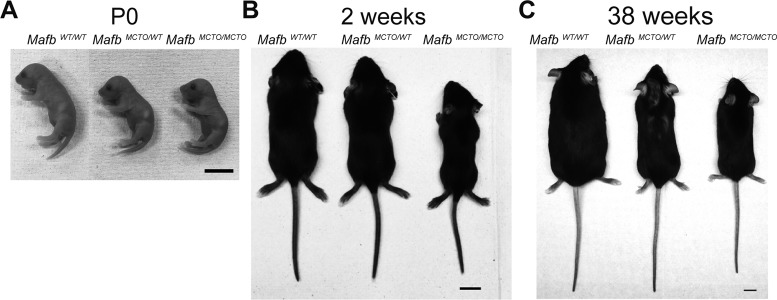
Multicentric carpotarsal osteolysis (MCTO) is a condition involving progressive osteolysis of the carpal and tarsal bones that is associated with glomerular sclerosis and renal failure (MCTO nephropathy). Previous work identified an autosomal dominant missense mutation in the transactivation domain of the transcription factor MAFB as the cause of MCTO. Several methods are currently used for MCTO nephropathy treatment, but these methods are invasive and lead to severe side effects, limiting their use. Therefore, the development of alternative treatments for MCTO nephropathy is required; however, the pathogenesis of MCTO in vivo is unclear without access to a mouse model. Here, we report the generation of an MCTO mouse model using the CRISPR/Cas9 system. These mice exhibit nephropathy symptoms that are similar to those observed in MCTO patients. MafbMCTO/MCTO mice show developmental defects in body weight from postnatal day 0, which persist as they age. They also exhibit high urine albumin creatinine levels from a young age, mimicking the nephropathic symptoms of MCTO patients. Characteristics of glomerular sclerosis reported in human patients are also observed, such as histological evidence of focal segmental glomerulosclerosis (FSGS), podocyte foot process microvillus transformation and podocyte foot process effacement. Therefore, this study contributes to the development of an alternative treatment for MCTO nephropathy by providing a viable mouse model.

Keywords: MafB; focal segmental glomerulosclerosis; multicentric carpotarsal osteolysis.


Figures

Fig. 1.
Fig. 1.
Generation of MafbMCTO/MCTO mice using the CRISPR/Cas9 system. (A) The upper sequence shows the Mafb domains within the genome. Red box, transactivation domain; green box, extended homology region; blue box, basic region; gray box, leucine zipper domain. The middle sequence shows the CRISPR target sequence at position 169–189 bp of the Mafb gene (boxed). The 200-nt oligo DNA sequence is shown in the lower sequence. The red bold face C/G in the middle sequence and T in the lower sequence indicate the target nucleotide mutation. (B) Genomic sequence of the CRISPR target site in MafbWT/WT and MafbMCTO/MCTO mice. Bold letters in the black box indicate the target mutation. (C) Genotyping methods for MafbMCTO/MCTO mice. In the upper sequence, the MafbWT allele and its associated primer are illustrated (Mafb mut genotype wtF). The 176C>T mutation and its associated primer are shown in the lower sequence (Mafb mut genotype mutF). The genotype of each mouse was determined via PCR. Representative results of PCR are shown in the lower images.
Fig. 2.
Fig. 2.
The MCTO mutation influences subsequent body growth. (A) Representative photographs of MafbWT/WT, MafbMCTO/WT, and MafbMCTO/MCTO littermates at P0. Scale bar: 1 cm. (B) Representative photographs of MafbWT/WT, MafbMCTO/WT, and MafbMCTO/MCTO littermates at 2 weeks. Scale bar: 1 cm. (C) Representative photographs of MafbWT/WT, MafbMCTO/WT , and MafbMCTO/MCTO littermates at 38 weeks. Scale bar: 1 cm. Table 1 provides the average weights of each genotype from P0 to 38 weeks. Supplementary Table 1, 2 and 3 provides the body weights of individual mice. The results for P0 and 2 weeks are from both male and female mice. Measurements from 2 weeks onwards were performed only in female mice. The data are presented as the mean ± SEM.; *P<0.05 (Student’s t-test); **P<0.01 (Student’s t-test).
Fig. 3.
Fig. 3.
MafbMCTO/MCTO mice exhibit an increased urine albumin creatinine ratio and FSGS phenotypes. (A) Urinary albumin creatinine ratios measured in MafbWT/WT, MafbMCTO/WT, and MafbMCTO/MCTO mice from 4 weeks to 38 weeks. The sample numbers of each genotype were as follows (for MafbWT/WT, MafbMCTOT/WT, and MafbMCTO/MCTO, respectively): 3, 4, and 3 at 4 weeks; 3, 4, and 4 at 8 to 12 weeks; 3, 4, and 6 at 14 to 18 weeks; 6, 4, and 9 at 20 and 26 weeks; and 3, 4, and 6 at 24, 28, and 38 weeks. Measurements at 4 weeks were performed in both male and female mice, while measurements at other times were performed only in female mice. (B) Kidneys of MafbWT/WT and MafbMCTO/MCTO mice subjected to PAS staining. Representative images are shown. Scale bar: 100 µm (left). The ratio of sclerotic glomeruli to total glomeruli was calculated in a blinded test (MafbWT/WT, n=4; MafbMCTO/MCTO, n=3). Solid gray circles, MafbWT/WT; solid white circles, MafbMCTO/MCTO (right). (C) Kidneys of MafbWT/WT and MafbMCTO/MCTO mice stained with Masson’s trichrome staining. Representative images are shown. Scale bar: 100 µm. Yellow arrowhead, sclerotic regions of a single glomerulus; blue area, fibrotic regions. Histological analyses were conducted using 25- and 26-week-old mice. Supplementary Table 4, 5, and 6 provides the urine albumin creatinine ratios of individual mice. The data are presented as the mean ± SEM.; *P<0.05 (Student’s t-test); **P<0.01 (Student’s t-test).
Fig. 4.
Fig. 4.
MafbMCTO/MCTO mice exhibit podocyte foot process effacement and microvillous transformation. (A) Electron microscopy image of 26-week-old female MafbWT/WT and MafbMCTO/MCTO glomeruli. Representative image are shown. White arrowhead, glomerular basement membrane; black arrowhead, Bowman’s space; scale bar: 2 µm (MafbWT/WT, n=3; MafbMCTO/MCTO, n=3). (B) Electron microscopy images of 26-week-old female MafbWT/WT and MafbMCTO/MCTO podocyte foot processes at 5,000× magnification. Representative image are shown. PC, podocyte; GBM, glomerular basement membrane.

Similar articles

Cited by

References

    1. Aziz A., Soucie E., Sarrazin S., Sieweke M.H.2009. MafB/c-Maf deficiency enables self-renewal of differentiated functional macrophages. Science 326: 867–871. doi: 10.1126/science.1176056 - DOI - PubMed
    1. Connor A., Highton J., Hung N.A., Dunbar J., MacGinley R., Walker R.2007. Multicentric carpal-tarsal osteolysis with nephropathy treated successfully with cyclosporine A: a case report and literature review. Am. J. Kidney Dis. 50: 649–654. doi: 10.1053/j.ajkd.2007.06.014 - DOI - PubMed
    1. Cuevas V.D., Anta L., Samaniego R., Orta-Zavalza E., Vladimir de la Rosa J., Baujat G., Domínguez-Soto Á., Sánchez-Mateos P., Escribese M.M., Castrillo A., Cormier-Daire V., Vega M.A., Corbí Á.L.2017. MAFB Determines Human Macrophage Anti-Inflammatory Polarization: Relevance for the Pathogenic Mechanisms Operating in Multicentric Carpotarsal Osteolysis. J. Immunol. 198: 2070–2081. doi: 10.4049/jimmunol.1601667 - DOI - PubMed
    1. Escano C.S., Armando I., Wang X., Asico L.D., Pascua A., Yang Y., Wang Z., Lau Y.S., Jose P.A.2009. Renal dopaminergic defect in C57Bl/6J mice. Am. J. Physiol. Regul. Integr. Comp. Physiol. 297: R1660–R1669. doi: 10.1152/ajpregu.00147.2009 - DOI - PMC - PubMed
    1. Eychène A., Rocques N., Pouponnot C.2008. A new MAFia in cancer. Nat. Rev. Cancer 8: 683–693. doi: 10.1038/nrc2460 - DOI - PubMed

MeSH terms

Supplementary concepts