

ENPP1-Fc prevents neointima formation in generalized arterial calcification of infancy through the generation of AMP
- PMID: 30369595
- PMCID: PMC6204430
- DOI: 10.1038/s12276-018-0163-5
ENPP1-Fc prevents neointima formation in generalized arterial calcification of infancy through the generation of AMP
Abstract
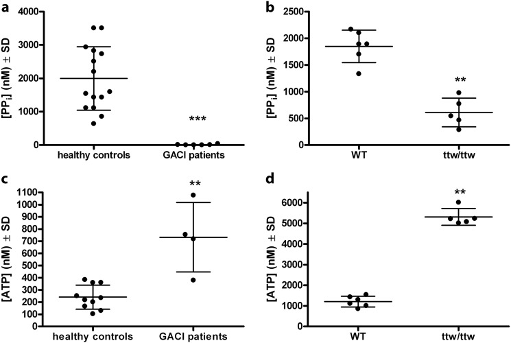
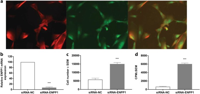
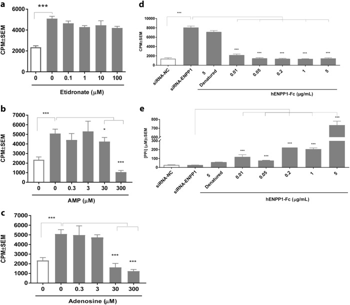
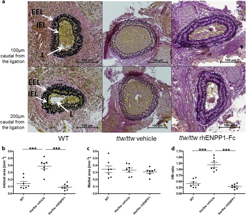
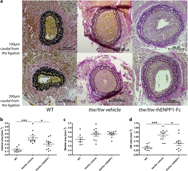
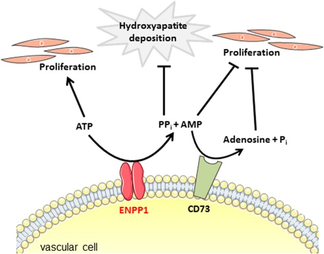
Generalized arterial calcification of infancy (GACI) is associated with widespread arterial calcification and stenoses and is caused by mutations in ENPP1. ENPP1 encodes for ectonucleotide pyrophosphatase/phosphodiesterase 1 (ENPP1), which cleaves ATP to generate inorganic pyrophosphate (PPi) and adenosine monophosphate (AMP) extracellularly. The current study was designed to define the prevalence of arterial stenoses in GACI individuals and to identify the mechanism through which ENPP1 deficiency causes intimal proliferation. Furthermore, we aimed to effectively prevent and treat neointima formation in an animal model of GACI through the systemic administration of recombinant human (rh)ENPP1-Fc protein. Based on a literature review, we report that arterial stenoses are present in at least 72.4% of GACI cases. We evaluated the effect of rhENPP1-Fc on ENPP1-silenced human vascular smooth muscle cells (VSMCs) and on induced intimal proliferation in Enpp1-deficient ttw/ttw mice treated with carotid ligation. We demonstrate that silencing ENPP1 in VSMCs resulted in a tenfold increase in proliferation relative to that of cells transfected with negative control siRNA. The addition of rhENPP1-Fc, AMP or adenosine restored the silenced ENPP1-associated proliferation. In contrast, neither PPi nor etidronate, a current off-label treatment for GACI, had an effect on VSMC proliferation. Furthermore, subcutaneous rhENPP1-Fc protein replacement was effective in preventing and treating intimal hyperplasia induced by carotid ligation in an animal model of GACI. We conclude that ENPP1 inhibits neointima formation by generating AMP. RhENPP1-Fc may serve as an approach for the effective prevention and treatment of arterial stenoses in GACI.
Conflict of interest statement
Y.Y. and K.A. were employees of Alexion Pharmaceuticals, Inc. at the time of the study. The company holds a patent on recombinant ENPP1-Fc. The remaining authors declare that they have no conflict of interest.
Figures
Similar articles
-
Inhibition of Vascular Smooth Muscle Cell Proliferation by ENPP1: The Role of CD73 and the Adenosine Signaling Axis.Cells. 2024 Jun 29;13(13):1128. doi: 10.3390/cells13131128. Cells. 2024. PMID: 38994980 Free PMC article.
-
Mono-allelic and bi-allelic ENPP1 deficiency promote post-injury neointimal hyperplasia associated with increased C/EBP homologous protein expression.Atherosclerosis. 2014 Apr;233(2):493-502. doi: 10.1016/j.atherosclerosis.2014.01.003. Epub 2014 Jan 21. Atherosclerosis. 2014. PMID: 24530784 Free PMC article.
-
ENPP1 enzyme replacement therapy improves blood pressure and cardiovascular function in a mouse model of generalized arterial calcification of infancy.Dis Model Mech. 2018 Oct 8;11(10):dmm035691. doi: 10.1242/dmm.035691. Dis Model Mech. 2018. PMID: 30158213 Free PMC article.
-
ENPP1 in Blood and Bone: Skeletal and Soft Tissue Diseases Induced by ENPP1 Deficiency.Annu Rev Pathol. 2024 Jan 24;19:507-540. doi: 10.1146/annurev-pathmechdis-051222-121126. Epub 2023 Oct 23. Annu Rev Pathol. 2024. PMID: 37871131 Free PMC article. Review.
-
Generalized Arterial Calcification of Infancy: New Insights, Controversies, and Approach to Management.Curr Osteoporos Rep. 2020 Jun;18(3):232-241. doi: 10.1007/s11914-020-00577-4. Curr Osteoporos Rep. 2020. PMID: 32172442 Free PMC article. Review.
Cited by
-
Longitudinal assessment of vascular calcification in generalized arterial calcification of infancy.Pediatr Radiol. 2022 Nov;52(12):2329-2341. doi: 10.1007/s00247-022-05364-0. Epub 2022 Apr 19. Pediatr Radiol. 2022. PMID: 35438330 Free PMC article. Review.
-
Generalized Arterial Calcification of Infancy Mimicking Coarctation of Aorta in a Neonate.Radiol Cardiothorac Imaging. 2024 Jun;6(3):e230403. doi: 10.1148/ryct.230403. Radiol Cardiothorac Imaging. 2024. PMID: 38900025 Free PMC article.
-
Improving the Pharmacodynamics and In Vivo Activity of ENPP1-Fc Through Protein and Glycosylation Engineering.Clin Transl Sci. 2021 Jan;14(1):362-372. doi: 10.1111/cts.12887. Epub 2020 Oct 20. Clin Transl Sci. 2021. PMID: 33064927 Free PMC article.
-
Weighing the Evidence for the Roles of Plasma Versus Local Pyrophosphate in Ectopic Calcification Disorders.J Bone Miner Res. 2023 Apr;38(4):457-463. doi: 10.1002/jbmr.4791. Epub 2023 Mar 13. J Bone Miner Res. 2023. PMID: 36807615 Free PMC article.
-
Disorders of phosphate homeostasis in children, part 1: primer on mineral ion homeostasis and the roles of phosphate in skeletal biology.Pediatr Radiol. 2022 Nov;52(12):2278-2289. doi: 10.1007/s00247-022-05374-y. Epub 2022 May 10. Pediatr Radiol. 2022. PMID: 35536415 Review.
References
Publication types
MeSH terms
Substances
Supplementary concepts
LinkOut - more resources
Full Text Sources
Other Literature Sources
Miscellaneous
