. 2018 Sep-Oct;51(5):321-327.
doi: 10.1590/0100-3984.2016.0134.

Idiopathic interstitial pneumonias: review of the latest American Thoracic Society/European Respiratory Society classification

Affiliations

Idiopathic interstitial pneumonias: review of the latest American Thoracic Society/European Respiratory Society classification

Daniel Simões Oliveira et al. Radiol Bras. 2018 Sep-Oct.

Abstract

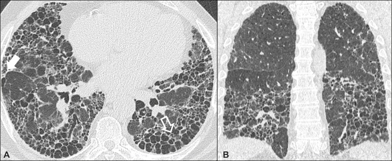
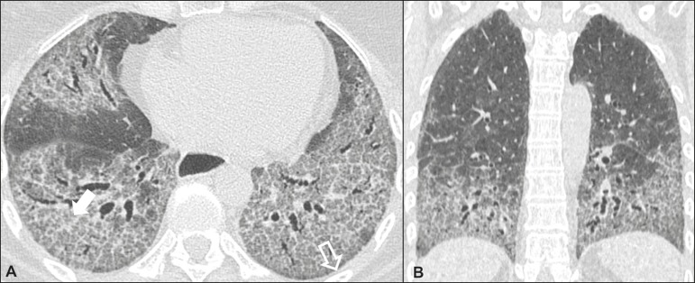
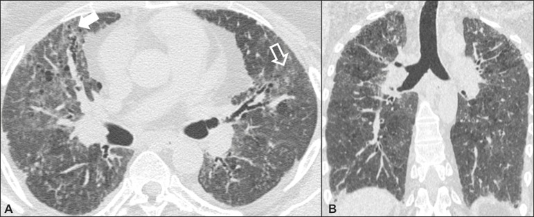
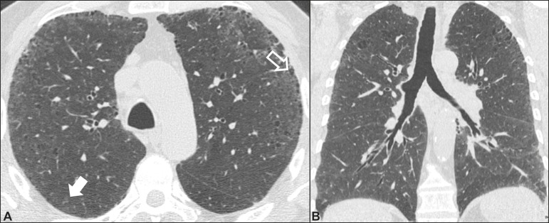
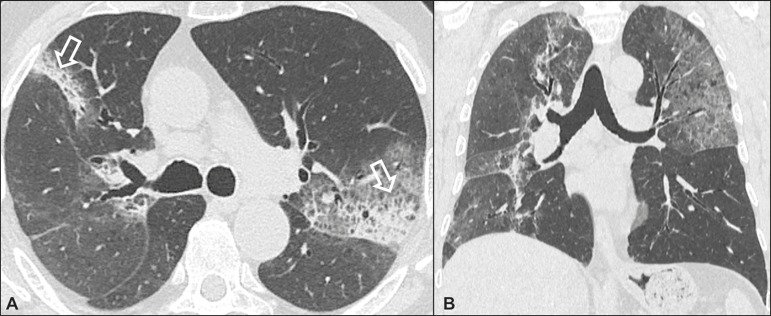
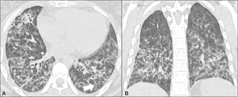
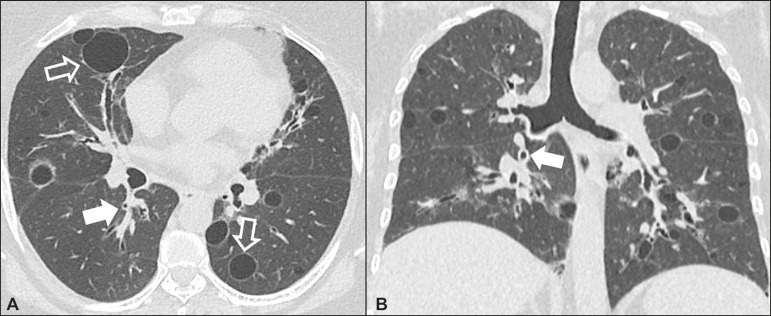
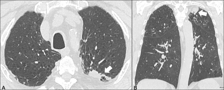
The diagnosis of idiopathic interstitial pneumonias (IIPs) involves a multidisciplinary scenario in which the radiologist assumes a key role. The latest (2013) update of the IIP classification by the American Thoracic Society/European Respiratory Society proposed some important changes to the original classification of 2002. The novelties include the addition of a new disease (idiopathic pleuroparenchymal fibroelastosis) and the subdivision of the IIPs into four main groups: chronic fibrosing IIPs (idiopathic pulmonary fibrosis and nonspecific interstitial pneumonia); smoking-related IIPs (desquamative interstitial pneumonia and respiratory bronchiolitis-associated interstitial lung disease); acute or subacute IIPs (cryptogenic organizing pneumonia and acute interstitial pneumonia); rare IIPs (lymphoid interstitial pneumonia and idiopathic pleuroparenchymal fibroelastosis); and the so-called "unclassifiable" IIPs. In this study, we review the main clinical, tomographic, and pathological characteristics of each IIP.

ResumoO diagnóstico das pneumonias intersticiais idiopáticas (PIIs) envolve um cenário multidisciplinar no qual o radiologista assume papel fundamental. A última atualização (2013) da classificação das PIIs pela American Thoracic Society/European Respiratory Society propõe algumas mudanças importantes em relação à classificação original de 2002. Dentre as novidades, destacam-se o acréscimo de uma nova doença (fibroelastose pleuroparenquimatosa idiopática) e a subdivisão das PIIs em quatro grupos principais: PIIs crônicas fibrosantes (fibrose pulmonar idiopática e pneumonia intersticial não específica); PIIs relacionadas ao tabagismo (pneumonia intersticial descamativa e bronquiolite respiratória com doença intersticial pulmonar); PIIs agudas/subagudas (pneumonia em organização e pneumonia intersticial aguda); PIIs raras (pneumonia intersticial linfocítica e fibroelastose pleuroparenquimatosa idiopática); além das ditas “inclassificáveis”. Foram revisadas, de forma didática neste estudo, as principais características clínicas, tomográficas e patológicas de cada uma das PIIs.

Keywords: American Thoracic Society; European Respiratory Society; Idiopathic interstitial pneumonia.


Figures

Figure 1
Figure 1
Idiopathic pulmonary fibrosis. A 66-year-old male smoker. Axial high-resolution computed tomography scan of the chest (A) and coronal reformatting (B). In A, fine reticular opacities (closed arrow), with traction bronchiectasis, traction bronchiolectasis, and honeycombing (open arrow). In B, predominantly peripheral and basal pattern of distribution.
Figure 2
Figure 2
Nonspecific interstitial pneumonia. A 51-year-old female patient with scleroderma. Axial high-resolution computed tomography scan of the chest (A) and coronal reformatting (B). In A, ground-glass attenuation, with linear reticular opacities (closed arrow), traction bronchiectasis, and traction bronchiolectasis. Note the discrete subpleural preservation (open arrow). In B, predominantly basal and symmetric pattern of distribution.
Figure 3
Figure 3
Desquamative interstitial pneumonia. A 65-year-old male smoker. Axial high-resolution computed tomography scan of the chest (A) and coronal reformatting (B). In A, linear reticular opacities (closed arrow), with sparse bilateral ground-glass opacity (open arrow). In B, predominantly peripheral pattern of distribution.
Figure 4
Figure 4
Respiratory bronchiolitis-associated interstitial lung disease. A 53-year-old male smoker. Axial high-resolution computed tomography scan of the chest (A) and coronal reformatting (B). In A, diffuse centrilobular opacities (closed arrow), thickening of the bronchial walls and paraseptal/centrilobular emphysema (open arrow). In B, distribution predominantly in the upper fields.
Figure 5
Figure 5
Cryptogenic organizing pneumonia. A 62-year-old female patient with a one-year history of dyspnea and a two-month history of symptom worsening. Axial high-resolution computed tomography scan of the chest (A) and coronal reformatting (B). In A, ground-glass opacities/sparse bilateral consolidations (open arrows). In B, bilateral subpleural and peribronchial pattern of distribution.
Figure 6
Figure 6
Acute interstitial pneumonia. A 32-year-old female patient, hospitalized for severe dyspnea two weeks prior. Axial high-resolution computed tomography scan of the chest (A) and coronal reformatting (B). In A, diffuse ground-glass attenuation pattern, with some traction bronchiectasis (open arrow) and reticular opacities (closed arrow). In B, bilateral diffuse, symmetrical pattern of distribution.
Figure 7
Figure 7
Lymphocytic interstitial pneumonia. A 62-year-old female patient with Sjögren’s syndrome. Axial high-resolution computed tomography scan of the chest (A) and coronal reformatting (B). In A, diffuse thickening of the bronchial walls (closed arrows), some ground-glass opacities and thin-walled cysts of varying sizes, with a diffuse, bilateral distribution (open arrows). In B, distribution predominantly in the lower fields.
Figure 8
Figure 8
Idiopathic pleuroparenchymal fibroelastosis. An 80-year-old male smoker with a one-year history of dyspnea on minimal exertion. Axial high-resolution computed tomography scan of the chest (A) and coronal reformatting (B). In A, pleural thickening with subpleural fibrotic changes (closed arrows). In B, asymmetric peripheral pattern of distribution in the upper fields.

References

    1. Torres PPTS, Moreira MAR, Silva DGST, et al. High-resolution computed tomography and histopathological findings in hypersensitivity pneumonitis: a pictorial essay. Radiol Bras. 2016;49:112–116. - PMC - PubMed
    1. Mogami R, Goldenberg T, Marca PGC, et al. Pulmonary infection caused by Mycobacterium kansasii: findings on computed tomography of the chest. Radiol Bras. 2016;49:209–213. - PMC - PubMed
    1. Queiroz RM, Gomes MP, Valentin MVN. Pulmonary paracoccidioidomycosis showing reversed halo sign with nodular/coarse contour. Radiol Bras. 2016;49:59–60. - PMC - PubMed
    1. Koenigkam-Santos M, Cruvinel DL, Menezes MB, et al. Quantitative computed tomography analysis of the airways in patients with cystic fibrosis using automated software: correlation with spirometry in the evaluation of severity. Radiol Bras. 2016;49:351–357. - PMC - PubMed
    1. Bastos AL, Corrêa RA, Ferreira GA. Tomography patterns of lung disease in systemic sclerosis. Radiol Bras. 2016;49:316–321. - PMC - PubMed

LinkOut - more resources