. 2018 Oct 9:9:2341.
doi: 10.3389/fmicb.2018.02341. eCollection 2018.

The Mycobacterial Cell Envelope: A Relict From the Past or the Result of Recent Evolution?

Affiliations

The Mycobacterial Cell Envelope: A Relict From the Past or the Result of Recent Evolution?

Antony T Vincent et al. Front Microbiol. .

Abstract

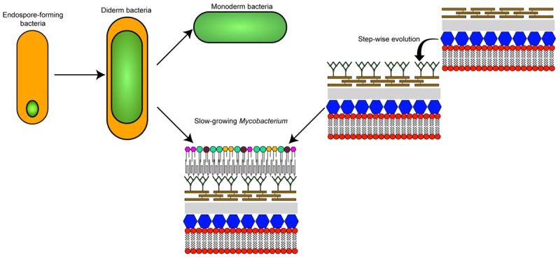
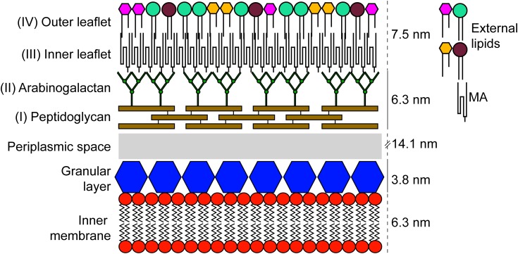
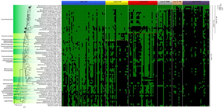
Mycobacteria are well known for their taxonomic diversity, their impact on global health, and for their atypical cell wall and envelope. In addition to a cytoplasmic membrane and a peptidoglycan layer, the cell envelope of members of the order Corynebacteriales, which include Mycobacterium tuberculosis, also have an arabinogalactan layer connecting the peptidoglycan to an outer membrane, the so-called "mycomembrane." This unusual cell envelope composition of mycobacteria is of prime importance for several physiological processes such as protection from external stresses and for virulence. Although there have been recent breakthroughs in the elucidation of the composition and organization of this cell envelope, its evolutionary origin remains a mystery. In this perspectives article, the characteristics of the cell envelope of mycobacteria with respect to other actinobacteria will be dissected through a molecular evolution framework in order to provide a panoramic view of the evolutionary pathways that appear to be at the origin of this unique cell envelope. In combination with a robust molecular phylogeny, we have assembled a gene matrix based on the presence or absence of key determinants of cell envelope biogenesis in the Actinobacteria phylum. We present several evolutionary scenarios regarding the origin of the mycomembrane. In light of the data presented here, we also propose a novel alternative hypothesis whereby the stepwise acquisition of core enzymatic functions may have allowed the sequential remodeling of the external cell membrane during the evolution of Actinobacteria and has led to the unique mycomembrane of slow-growing mycobacteria as we know it today.

Keywords: Actinobacteria; Mycobacterium; cell envelope; evolution; genomics.


Figures

GRAPHICAL ABSTRACT
GRAPHICAL ABSTRACT
The different theories on the evolutionary origin of the mycobacterial cell envelope.
FIGURE 1
FIGURE 1
Schematic representation of the cell envelope found in members of the order Corynebacteriales. The components were scaled up using results obtained by CEMOVIS for M. bovis BCG (Zuber et al., 2008).
FIGURE 2
FIGURE 2
Distribution within the Actinobacteria phylum of genes involved in the synthesis of components of the cell envelope of M. tuberculosis. The 72 genome sequences (Supplementary Table S1), comprising at least one species per order, were annotated using PROKKA version 1.12 (Seemann, 2014). The phylogenetic tree was generated from 86 sequences inferred as orthologous by GET_HOMOLOGUES version 20180313 (Contreras-Moreira and Vinuesa, 2013). The sequences were aligned using MAFFT version 7.397 (Katoh and Standley, 2013) and concatenated and partitioned using AMAS (Borowiec, 2016). Finally, the best evolutionary model for each partition was found by IQ-TREE version 1.6.3 (Kalyaanamoorthy et al., 2017) and maximum-likelihood phylogenetic analysis was also performed using IQ-TREE (Nguyen et al., 2015) using 10,000 ultrafast bootstrap replicates (Hoang et al., 2018). Only bootstrap values under 100 are shown on the tree. Nodes that are discussed in the article are highlighted with a numbered black circle. The green and black squares of the matrix highlight genes that are present or absent, respectively. Individual genes (Supplementary Table S2) were considered to be present when they had a sequence similarity ≥50% relative to M. tuberculosis [an e-value cut-off of 1e-10 has also been applied in TBLASTN version 2.7.1 (Altschul et al., 1997)]. The raw data, containing the similarity values, is presented in the Supplementary Table S2 for clarity. The “” represent micro-evolution of specific strains (described in the corresponding sections of the article). Dashed lines represent delineation, described in the literature, of the presence/absence of the specific layers also depicted at the right side of the figure. Of note, although the phylogenetic position of Gaiella sp. SCGC AG-212-M14 is expected for a member of the order Gaiellales, the size of the assembly (<1 Mb) coming from a single cell amplified by WGA-X, suggests that the sequence of this strain is likely incomplete.

References

    1. Altschul S. F., Madden T. L., Schaffer A. A., Zhang J., Zhang Z., Miller W., et al. (1997). Gapped BLAST and PSI-BLAST: a new generation of protein database search programs. Nucleic Acids Res. 25 3389–3402. - PMC - PubMed
    1. Baek I., Kim M., Lee I., Na S. I., Goodfellow M., Chun J. (2018). Phylogeny trumps chemotaxonomy: a case study involving Turicella otitidis. Front. Microbiol. 9:834. 10.3389/fmicb.2018.00834 - DOI - PMC - PubMed
    1. Bansal-Mutalik R., Nikaido H. (2014). Mycobacterial outer membrane is a lipid bilayer and the inner membrane is unusually rich in diacyl phosphatidylinositol dimannosides. Proc. Natl. Acad. Sci. U.S.A. 111 4958–4963. 10.1073/pnas.1403078111 - DOI - PMC - PubMed
    1. Barka E. A., Vatsa P., Sanchez L., Gaveau-Vaillant N., Jacquard C., Meier-Kolthoff J. P., et al. (2016). Taxonomy, physiology, and natural products of Actinobacteria. Microbiol. Mol. Biol. Rev. 80 1–43. 10.1128/MMBR.00019-15 - DOI - PMC - PubMed
    1. Barksdale L., Kim K. S. (1977). Mycobacterium. Bacteriol. Rev. 41 217–372. - PMC - PubMed