Case Reports
. 2018 Jan;6(1):e70-e76.
doi: 10.1055/s-0038-1672165. Epub 2018 Oct 18.

Three-Dimensional Printed Model and Virtual Reconstruction: An Extra Tool for Pediatric Solid Tumors Surgery

Affiliations
Case Reports

Three-Dimensional Printed Model and Virtual Reconstruction: An Extra Tool for Pediatric Solid Tumors Surgery

Ángela Sánchez-Sánchez et al. European J Pediatr Surg Rep. 2018 Jan.

Abstract

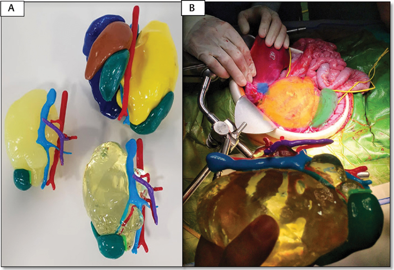
Introduction Three-dimensional (3D) technology is increasingly applied for planning challenging surgical interventions. We report our experience using 3D printing and virtual reconstruction for surgical planning of complex tumor resections in children. Methods Data were obtained from preoperative magnetic resonance. imaging analysis and 3D virtual recreations were performed using specialized computer software. 3D real-scale geometry models, including tumor, adjacent organs, and relevant vascularization, were printed in colorimetric scale and different materials for optimal structures discrimination. Results Four complex cases were selected. The first case was a bilateral Wilms tumor. The volumetric reconstruction proved the presence of enough healthy renal tissue, allowing bilateral nephron-sparing surgery. In the second case, reconstruction contributed to the location of pulmonary metastases. The third case was an abdominal neuroblastoma stage L2. The 3D model was of high value for planning and as a reference during the intervention. The last case is a cervico-thoracic neuroblastoma with an anatomopathological diagnosis of ganglioneuroma, located at the cervico-mediastinal juncture, in close relationship with the cervical vessels. Conclusions 3D reconstruction and the full-scale printing models are a useful tool in cases of complex tumor resections as they contribute to a better understanding of the relationships between the tumor and adjacent organs, helping to anticipate certain surgical complications. They also provide additional information to conventional imaging tests, being able to influence therapeutic decisions and facilitate the understanding by the family, improving doctor-patient communication.

Keywords: 3D printing; oncology; pediatrics; stereolithography; virtual reconstruction.


Conflict of interest statement

Conflict of Interest None.

Figures

Fig. 1
Fig. 1
Different steps of 3D printing are shown. ( A ) FDM printer; ( B ) printed structure still with support material. ( C ) Result after post-processing the piece. FDM, fused deposit modeling; 3D, three-dimensional.
Fig. 2
Fig. 2
Case 1. ( A ) Coronal MRI section showing both kidneys affected by Wilms tumor, confirming the large size of the left tumor mass. ( B and C ) Volumetric reconstructions of both tumors. Healthy renal parenchyma susceptible to nephron sparing surgery was proved. LK, left kidney; MRI, magnetic resonance imaging; RK, right kidney.
Fig. 3
Fig. 3
Case 1. The images ( AC ) show different moments of the surgical intervention. After bilateral nephron-sparing surgery, coagulation was performed with adhesive matrix and plication of the renal parenchyma. In the image ( D ), the resected specimens are shown together with their corresponding three-dimensional models, objectifying the great similarity between both.
Fig. 4
Fig. 4
Case 2. ( A ) Three-dimensional reconstruction performed from the preoperative CT. There are three metastases in the right lung and one in the left one. ( B and C ) Cross-sections of the thoracic CT. Lesions marked by arrow correspond to those marked with the same color in the virtual reconstruction. CT, computed tomography.
Fig. 5
Fig. 5
Case 3. ( A ) MRI showing a large abdominal mass that moves the liver upward. ( B ) Virtual reconstruction of the tumor (yellow), liver (green), both kidneys (gray), and spleen (brown). The involved vasculature was also reconstructed (aorta in red, vena cava in blue, and portal vein in purple). ( C ) Modified virtual reconstruction, observing the tumor of semitransparent consistency, which allows the visualization of the renal artery and vein within the tumor. The kidney is displaced inferiorly with loss of its morphology due to the mass effect. MRI, magnetic resonance imaging.
Fig. 6
Fig. 6
Case 3. ( A ) Different printed models representing the abdominal neuroblastoma. The superior model includes tumor (yellow), aorta (red) and vena cava (blue) with its respective branches, liver (blue), both kidneys (green), and spleen (brown). The different structures are assembled by a magnetization system. The other two models represent the tumor and the relevant vascularity in different textures. ( B ) The model was consulted several times during the surgery. In the image, the correspondence between the model and the surgical anatomy is observed.
Fig. 7
Fig. 7
Case 4. Virtual reconstruction of cervicothoracic paravertebral ganglioneuroma and comparison with images obtained by MRI (A-B coronal view, C-D transverse view). The tumor displaces the left carotid artery, the internal jugular vein, and the airway. The vertebral artery is encased by the tumor. MRI, magnetic resonance imaging.

References

    1. Souzaki R, Kinoshita Y, Ieiri S et al.Preoperative surgical simulation of laparoscopic adrenalectomy for neuroblastoma using a three-dimensional printed model based on preoperative CT images. J Pediatr Surg. 2015;50(12):2112–2115. - PubMed
    1. Krauel L, Fenollosa F, Riaza L et al.Use of 3D prototypes for complex surgical oncologic cases. World J Surg. 2016;40(04):889–894. - PubMed
    1. Zhao J, Zhou X J, Zhu C Z et al.3D simulation assisted resection of giant hepatic mesenchymal hamartoma in children. Comput Assist Surg (Abingdon) 2017;22(01):54–59. - PubMed
    1. Fuchs I, Tutschek B, Henrich W. Visualization of the fetal fontanels and skull sutures by three-dimensional translabial ultrasound during the second stage of labor. Ultrasound Obstet Gynecol. 2008;31(04):484–486. - PubMed
    1. Vijayavenkataraman S, Fuh J YH, Lu W F. 3D Printing and 3D bioprinting in pediatrics. Bioengineering (Basel) 2017;4(03):E63. - PMC - PubMed

Publication types