Review
. 2019 Jan;37(1):35-50.
doi: 10.1002/jor.24170. Epub 2018 Nov 30.

Cellular biology of fracture healing

Affiliations
Review

Cellular biology of fracture healing

Chelsea S Bahney et al. J Orthop Res. 2019 Jan.

Abstract

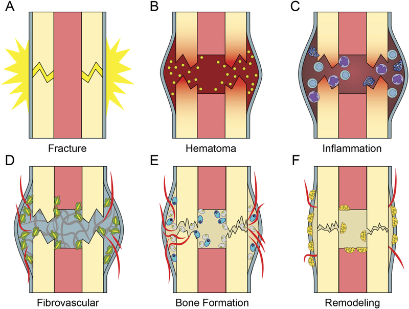
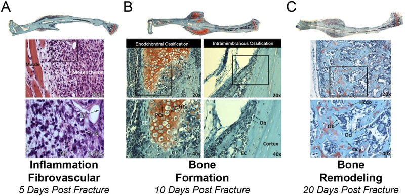
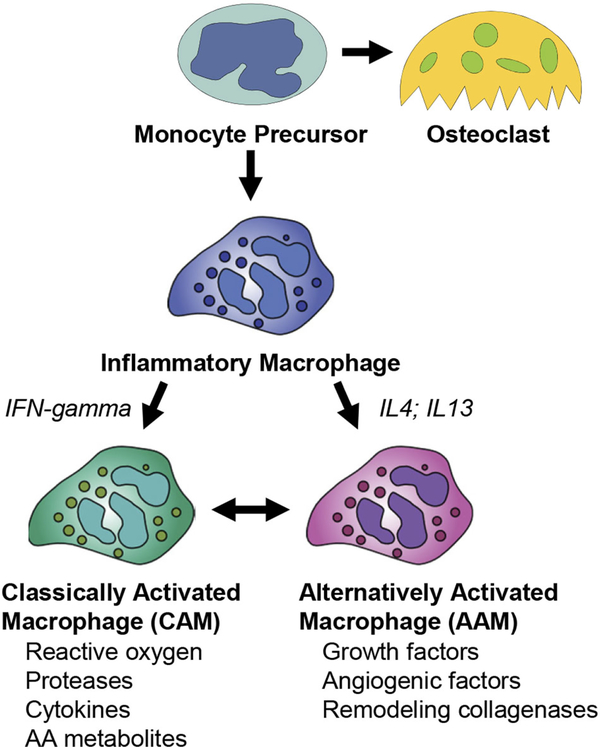
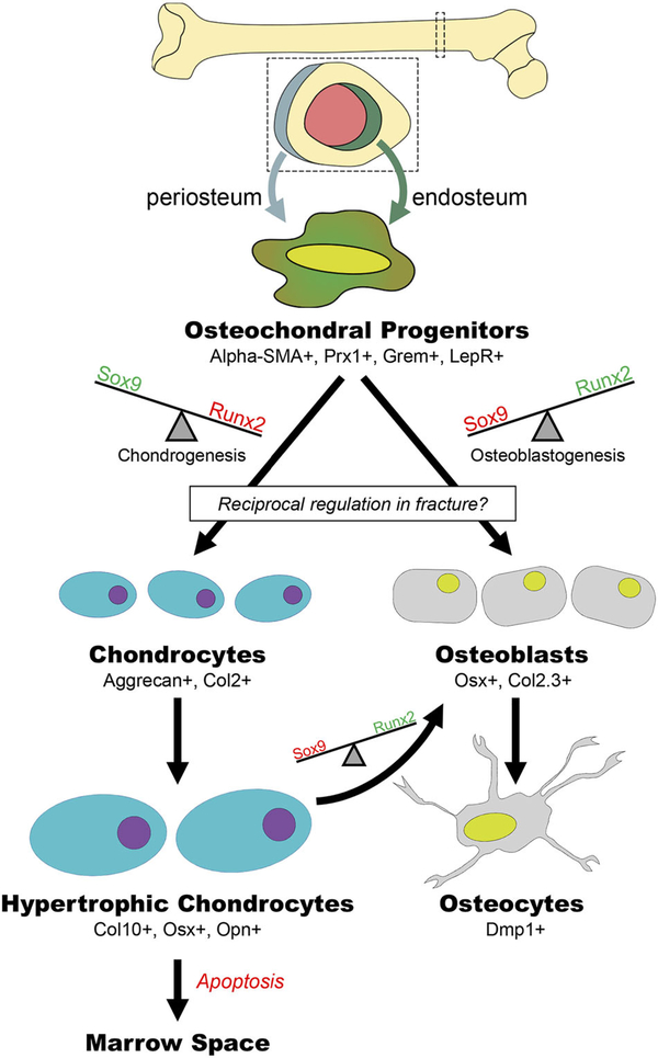
The biology of bone healing is a rapidly developing science. Advances in transgenic and gene-targeted mice have enabled tissue and cell-specific investigations of skeletal regeneration. As an example, only recently has it been recognized that chondrocytes convert to osteoblasts during healing bone, and only several years prior, seminal publications reported definitively that the primary tissues contributing bone forming cells during regeneration were the periosteum and endosteum. While genetically modified animals offer incredible insights into the temporal and spatial importance of various gene products, the complexity and rapidity of healing-coupled with the heterogeneity of animal models-renders studies of regenerative biology challenging. Herein, cells that play a key role in bone healing will be reviewed and extracellular mediators regulating their behavior discussed. We will focus on recent studies that explore novel roles of inflammation in bone healing, and the origins and fates of various cells in the fracture environment. © 2018 Orthopaedic Research Society. Published by Wiley Periodicals, Inc. J Orthop Res.

Keywords: bone regeneration; bone repair; fracture healing.


Figures

Figure 1.
Figure 1.
Fracture healing is temporally-defined process. (A) At injury there is disruption of periosteum and bone (B) A clot forms immediately providing a provisional matrix. Platelet degranulation releases chemokines to recruit inflammation. (C) Inflammatory phase leads to a period of (D) Mesenchymal expansion and migration from the periosteum and endosteum and angiogenesis, (E) Bone is formed via both endochondral (blue large oval cells) and intramembranous ossification (smaller grey cells), (F) Osteoclasts (multinucleated cells) resorb primary bone and the process of remodeling restores bone shape and structure.
Figure 2.
Figure 2.
Multiple cell-types present during the process of bone regeneration. Tibae were harvested 5 (A), 10 (B), and 20 (C) days post closed fracture and fixation with an intramedullary pin. Longitudinal histological sections were stained with H&E (A) or safranin-o (B and C) imaged at 2.5X and images stiched together and higher magnification images at 20X, 40X, and 100X obtained. (A) 5 day post-fracture undifferentiated mesenchymal cells are present in the callus and areas of inflammation remain (boxed area in 40X image is magnified in 100X) EC, endothelial cell; N, neutrophil; L, lymphocyte; M, macrophage. (B) 10 days post-fracture there is both endochondral ossification (red staining, safranin-o stains cartilage) and intramembranous bone formation occurring. Boxed areas in 20X images are magnified in 40X images. EC, endothelial cell; PC, proliferation chondrocytes; HC, hypertrophic chondrocytes; OB, osteoblast. (C) 20 days post-fracture. An extensive network of primary bone has formed and endochondral ossification is complete. Boxed areas in 20X images are magnified in 40X images. Ob, Osteoblast; Ocl, Osteoclast; Ocy, osteocyte; Hcdo, hypertrophic chondrocyte derived osteoblast.
Figure 3.
Figure 3.
Macrophage precursors develop into both classically activated and alternatively activated macrophages. Monocyte precursors give rise to both the osteoclast lineage and to inflammatory macrophages. Various factors, such as IFN-gamma, IL4, and IL13 control transitions between classically activated macrophages (CAM) and Alternatively activated macrophages (AAM).
Figure 4.
Figure 4.
Mesenchymal precursors develop into both osteoblasts and chondrocytes. Osteochondral progenitors are activated at the time of bone injury and a balance in transcriptional activation results in the cells becoming either osteoblasts or chondrocytes. Hypertrophic chondrocytes can differentiate to become osteoblasts.
Figure 5.
Figure 5.
Hypertrophic chondrocytes develop into osteoblasts and osteocytes. Tibiae were harvested post fracture and stained with (A) Safranin-O to define the chondrogenic front as outlined in panel (B). (B) shows zones of hypertrophic chondrocytes, transition zone, Bone, and blood vessels (BV). (C) is a low magnification H&E image showing the localization of panels D and E in areas of bone. Cells of bone can be traced to the chondrocyte lineage using the (D) Col2CreERT2:: Ai9 or the (E) AgcCreERT::Ai9 mouse with a tamoxifen pulse.

Similar articles

Cited by

References

    1. Wang Y, Wan C, Deng L, et al. 2007. The hypoxiainducible factor {alpha} pathway couples angiogenesis to osteogenesis during skeletal development. J Clin Invest 117:1616–1626. - PMC - PubMed
    1. Hu DP, Ferro F, Yang F, et al. 2017. Cartilage to bone transformation during fracture healing is coordinated by the invading vasculature and induction of the core pluripotency genes. Development 144:221–234. - PMC - PubMed
    1. Einhorn TA. 1995. Enhancement of fracture-healing. J Bone Joint Surg Am 77:940–956. - PubMed
    1. Barnes GL, Kostenuik PJ, Gerstenfeld LC, et al. 1999. Growth factor regulation of fracture repair. J Bone Miner Res 14:1805–1815. - PubMed
    1. Rapp AE, Bindl R, Recknagel S, et al. 2016. Fracture healing is delayed in immunodeficient NOD/scidIL2Rgammacnull mice. PLoS ONE 11:e0147465. - PMC - PubMed