. 2018 Sep 18;7(18):e009112.
doi: 10.1161/JAHA.118.009112.

Calorie Restriction Curbs Proinflammation That Accompanies Arterial Aging, Preserving a Youthful Phenotype

Affiliations

Calorie Restriction Curbs Proinflammation That Accompanies Arterial Aging, Preserving a Youthful Phenotype

Mingyi Wang et al. J Am Heart Assoc. .

Erratum in

Abstract

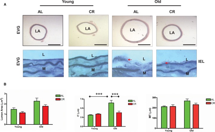
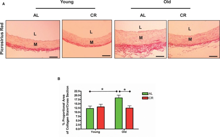
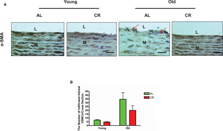
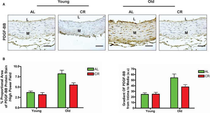
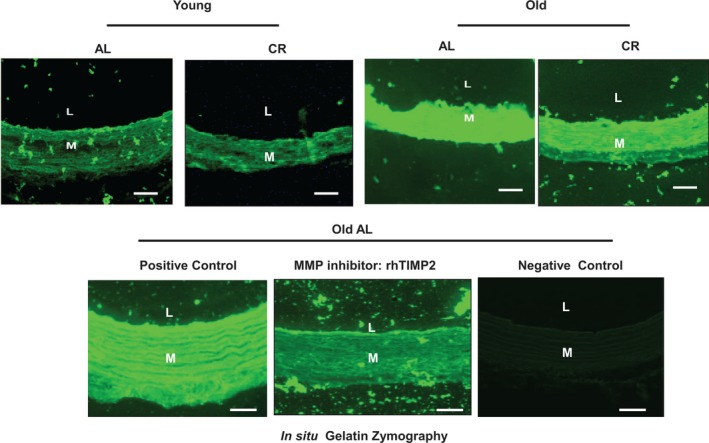
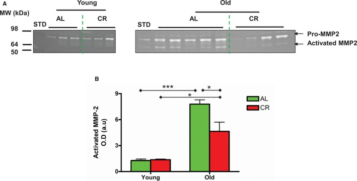
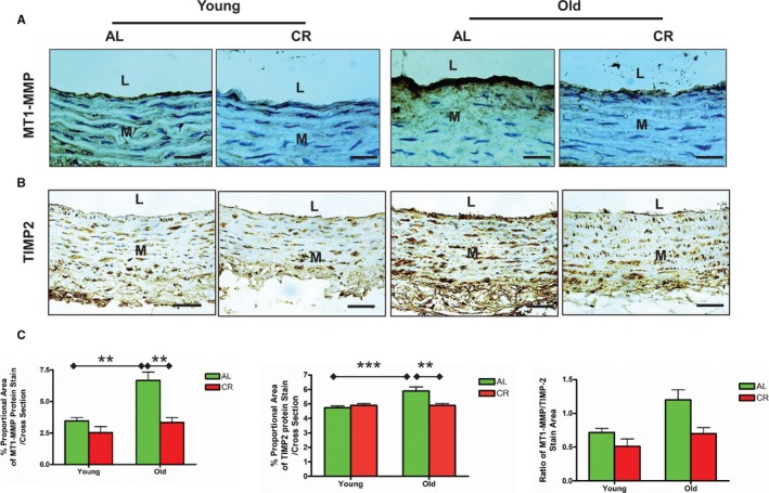
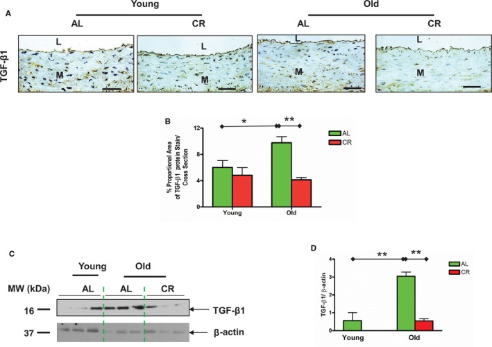
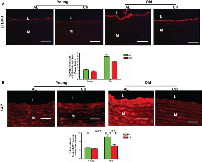
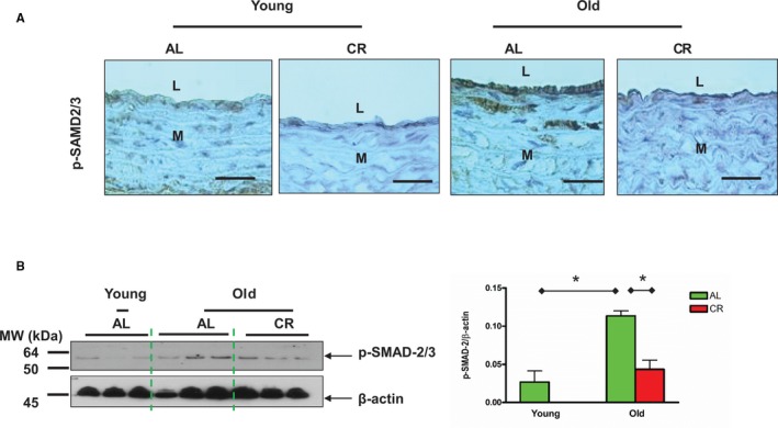
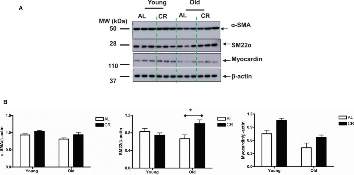
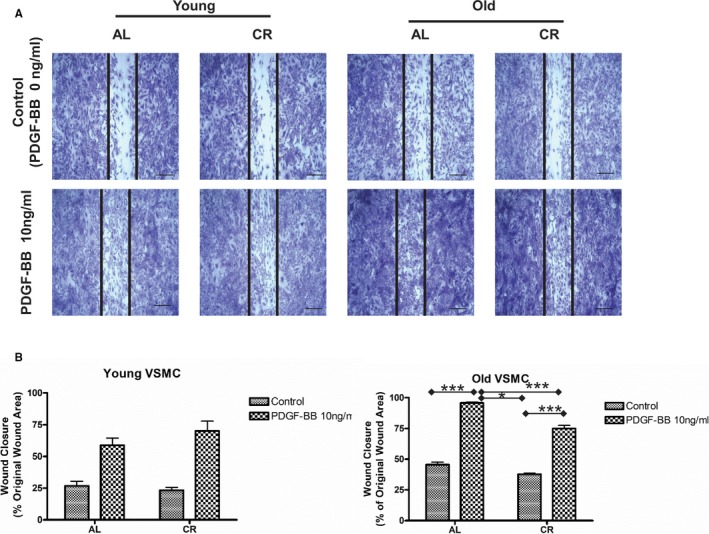
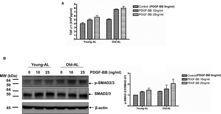
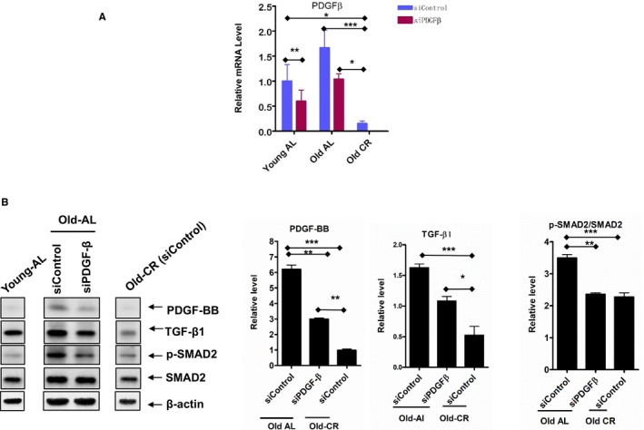
Background Aging exponentially increases the incidence of morbidity and mortality of quintessential cardiovascular disease mainly due to arterial proinflammatory shifts at the molecular, cellular, and tissue levels within the arterial wall. Calorie restriction ( CR ) in rats improves arterial function and extends both health span and life span. How CR affects the proinflammatory landscape of molecular, cellular, and tissue phenotypic shifts within the arterial wall in rats, however, remains to be elucidated. Methods and Results Aortae were harvested from young (6-month-old) and old (24-month-old) Fischer 344 rats, fed ad libitum and a second group maintained on a 40% CR beginning at 1 month of age. Histopathologic and morphometric analysis of the arterial wall demonstrated that CR markedly reduced age-associated intimal medial thickening, collagen deposition, and elastin fractionation/degradation within the arterial walls. Immunostaining/blotting showed that CR effectively prevented an age-associated increase in the density of platelet-derived growth factor, matrix metalloproteinase type II activity, and transforming growth factor beta 1 and its downstream signaling molecules, phospho-mothers against decapentaplegic homolog-2/3 (p- SMAD -2/3) in the arterial wall. In early passage cultured vascular smooth muscle cells isolated from AL and CR rat aortae, CR alleviated the age-associated vascular smooth muscle cell phenotypic shifts, profibrogenic signaling, and migration/proliferation in response to platelet-derived growth factor. Conclusions CR reduces matrix and cellular proinflammation associated with aging that occurs within the aortic wall and that are attributable to platelet-derived growth factor signaling. Thus, CR reduces the platelet-derived growth factor-associated signaling cascade, contributing to the postponement of biological aging and preservation of a more youthful aortic wall phenotype.

Keywords: aging; arterial remodeling; calorie restriction; proinflammation; rats; vascular smooth muscle cells.


Figures

Figure 1
Figure 1
CR retards age‐associated aortic wall remodeling. A, Representative photomicrographs of paraffin sections of thoracic aortae from both young and old F344 rats with an AL or a CR diet stained with EVG (upper panels: EVG stain elastin as dark blue, ×50); and representative photomicrographs of paraffin sections of thoracic aortae from both young and old Fischer 344 rats fed an AL or a CR diet stained with EVG (elastin laminae as dark blue, ×1000). Arrow indicates eroded or broken IEL. Bar scale, 200 μm. B, Data shown as mean±SEM (n=5 animals/group). Left panel: LA was analyzed with a 2‐way ANOVA showing no age×diet interaction (P>0.05), after removing this term from the model, the overall main age (P<0.05) and diet (P<0.01) effects were significant. Middle panel of IT was also done with a 2‐way ANOVA followed by a Bonferroni comparisons test: P<0.001 for age×diet interaction. Post hoc comparisons: old vs young within AL group and AL vs CR in old group were reported (***P<0.001). Right Panel of MT: In a 2‐way ANOVA, there were no age×diet interactions (P>0.05), but overall main age effect was reported (P<0.05). AL indicates ad libitum; CR, calorie restriction; EVG, Elastic‐van Gieson; IEL, intimal elastin laminae; IT, intimal thickness; L, lumen; LA, lumen area; M, media; MT, media thickness.
Figure 2
Figure 2
CR prevents age‐associated increases in elastin lamina fragmentation/degradation in the aortic walls. A, Representative photomicrographs of paraffin sections thoracic aortae from both young and old F344 rats with an AL or a CR diet stained with EVG (dark blue) (×400). Bar scale, 10 μm. B, Elastin fraction shown as mean±SEM (n=5 animals/group). The data were analyzed with a 2‐way ANOVA: P<0.01 for age×diet interaction. Pairwise comparisons: old vs young within AL group and AL vs CR in old group were shown (**P<0.01). AL indicates ad libitum; CR, calorie restriction; EVG, Elastica‐van Gieson; L, lumen; M, media.
Figure 3
Figure 3
CR retards age‐associated increases in collagen deposition within the aortic walls. A, Representative photomicrographs of paraffin thoracic aortic sections of both young and old F344 rats with an AL or a CR diet stained with Sirius Red for collagen (red), observed using conventional light microscopy (×200). n=5 rats/group, respectively. Bar scale, 20 μm. B, Collagen density shown as mean±SEM (n=5 animals/group). The data were analyzed with a 2‐way ANOVA: P<0.05 for age×diet interaction. Pairwise comparison: old vs young within AL group and AL vs CR in old group were shown (*P<0.05). AL indicates ad libitum; CR, calorie restriction; L, lumen; M, media.
Figure 4
Figure 4
CR affects age‐associated increases in VSMC infiltration in intima within the aortic walls. A, Representative photomicrographs of paraffin sections thoracic aortae from both young and old F344 rats with an AL or a CR diet, stained with an antibody against α‐SMA (brown, ×400) showing infiltrated intimal VSMCs (arrowhead). n=5 rats/group. Scale bar, 20 μm. B, The numbers of infiltrated VSMC presented as mean±SEM (n=5 animals/group). The data were analyzed with a 2‐way ANOVA and showed that there was no age×diet interaction (P>0.05); but an overall main age effect (P<0.001) was reported. AL indicates ad libitum; CR, calorie restriction; L, lumen; M, media; VSMC, vascular smooth muscle cells; α‐SMA, alpha‐smooth muscle actin.
Figure 5
Figure 5
CR inhibits PDGF expression within the aortic wall with aging. A, Representative of photomicrographs of immunostaining of PDGFBB on paraffin sections of the rat thoracic aortae (brown, upper panels, ×200). Scale bar, 10 μm. B, PDGFBB density or gradient shown as mean±SEM (n=3–5 animals/group). The data were analyzed with a 2‐way ANOVA and showed no age×diet interaction (P>0.05), but the overall main age effect (P<0.001) and diet effect (P<0.05) were reported in percent proportional area of PDGFBB and in the gradient of PDGFBB. AL indicates ad libitum; CR, calorie restriction; L, lumen; M, media; PDGF‐BB, platelet derived growth factor.
Figure 6
Figure 6
CR reduces aortic MMP‐2 activation in situ within the aortic wall with aging. Representative photomicrographs of in situ thoracic aortic gelatin zymogram from both young and old AL and CR F344 rats with or without MMP inhibitor: recombinant human TIMP2 (rhTIMP2) (500 ng/mL) (activated MMP, green color). Scale bar, 20 μm. AL indicates ad libitum; CR, calorie restriction; L, lumen; M, media; MMP, matrix metalloproteinase; rhTIMP2, recombinant human tissue inhibitor of metalloproteinases 2.
Figure 7
Figure 7
CR reduces aortic MMP‐2 activation in the aortic wall with aging. A, PAGE gelatin zymogram of lysates of intact thoracic aortae. B, Densitometric analysis of activated MMP‐2. Data shown as mean±SEM (n=3–5 animals/group). The data were analyzed with a 2‐way ANOVA showing a marginally significant age×diet interaction (P=0.06), age effect (P<0.001), and diet effect (P<0.001). Pairwise comparison: old vs young within AL or CR group, respectively, and AL vs CR in old group were reported (*P<0.05; ***P<0.001). AL indicates ad libitum; CR, calorie restriction; L, lumen; M, media; MMP‐2, matrix metalloproteinase type II.
Figure 8
Figure 8
CR decreases MT1‐MMP and TIMP2 protein expression within the aged aortic wall. A, Immunostaining for MT1‐MMP (×400). Bar scale, 10 μm. B, Immunostaining for TIMP2 (×400). Bar scale, 10 μm. C, Morphometric analysis for MT1‐MMP and TIMP2 immunostaining target to total aortic area ratio/cross section. Data shown as mean±SEM (n=5 animals/group). Left panel: The data of MT1‐MMP density was analyzed with a 2‐way ANOVA showing age×diet interaction (P<0.05). Pairwise comparisons (old vs young within AL group; AL vs CR in old group) were reported (**P<0.01). Middle panel: In 2‐way ANOVA of TIMP2 density, age×diet interaction (P<0.01) was found. Pairwise comparison: old vs young within AL group and AL vs CR in old group were reported (**P<0.01; ***P<0.001). Right panel: In a 2‐way ANOVA of the ratio of MT1‐MMP/TIMP2, age×diet interaction was not found (P>0.05), but overall main age effect (P<0.05) and diet effect (P<0.05) were reported. AL indicates ad libitum; CR, calorie restriction; L, lumen; M, media; MMP‐2, matrix metalloproteinase type II; MT1‐MMP, membrane type 1 matrix metalloproteinase; TIMP2, tissue inhibitor of metalloproteinases 2.
Figure 9
Figure 9
CR diminishes the age‐associated TGF‐β1 increase in the aortic wall. A, Immunostaining for TGF‐β1 (×400). Bar scale, 10 μm. B, Quantification of TGF‐β1 protein staining proportion shown mean±SEM (n=3 animals/group). Two‐way ANOVA followed by Bonferroni post hoc test showed that an age×diet interaction (P<0.05), pairwise comparison (old vs young within AL group; AL vs CR in old group) was reported (*P<0.05; **P<0.01). C, Immunoblotting TGF‐β1 from lysates of thoracic aortic tissue. D, Densitometric analysis of TGF‐β1. Data shown as mean±SEM (n=3 animals/group). One‐way ANOVA of TGF‐β1 abundance (P<0.05) followed by Bonferroni post hoc test, pairwise comparison (old vs young within AL group; AL vs CR in old group) was reported (**P<0.01). AL indicates ad libitum; CR, calorie restriction; L, lumen; M, media; TGF‐β1, transforming growth factor beta 1.
Figure 10
Figure 10
CR attenuates latent TGF‐β1 associated protein expression within aging aortic walls. A, Representative immunofluorescence staining for LTBP‐1 (upper panels, ×400, red) and quantification of the LTBP‐1 staining proportion (lower panel). Data shown as mean±SEM (n=3 animals/group). Two‐way ANOVA followed by Bonferroni post hoc test showed no age×diet interaction (P>0.05). Scale bar, 20 μm. B, Representative immunofluorescence staining for LAP (upper panels, ×400, red) and morphometric analysis of the LAP protein fraction. Data shown as mean±SEM (n=3 animals/group). Two‐way ANOVA followed by Bonferroni post hoc test, indicated an age×diet interaction was reported (P<0.05); pairwise comparison (old vs young within AL group; AL vs CR in old group) was presented (**P<0.01; ***P<0.001). Scale bar, 20 μm. AL indicates ad libitum; CR, calorie restriction; L, lumen; LAP, latent associated protein; LTBP‐1, latent TGF binding protein‐1; M, media.
Figure 11
Figure 11
CR decreases SMAD‐2/3 phosphorylation within aged aortic walls. A, Immunostaining for p‐SMAD‐2/3 (brown, upper panels, ×400). Scale bar, 20 μm. B, Immunoblotting for p‐SMAD‐2/3 (left side of panel). Densitometric analysis of p‐SMAD‐2/3 (right side). Data presented as mean±SEM (n=3 animals/group). One‐way ANOVA showed significance was found (P<0.05); pairwise comparison (old vs young within AL group; AL vs CR in old group) was presented (*P<0.05). AL indicates ad libitum; CR, calorie restriction; L, lumen; M, media; p‐SMAD‐2/3, phospho‐mothers against decapentaplegic homolog‐2/3.
Figure 12
Figure 12
CR modifies the phenotype of VSMCs isolated from old rats. A, Immunoblotting for α‐SMA, SM22α, and myocardin. B, Densitometric analysis. Data shown as mean±SEM (n=3–6 independent experiments). Left panel: α‐SMA protein abundance was analyzed with a 2‐way ANOVA followed by a Bonferroni comparisons test and showed no age×diet interaction (P>0.05), but an overall main age effect (P<0.05) and diet effect (P<0.05) were reported; Middle panel: In a 2‐way ANOVA of SM22α, an age×diet interaction was found (P<0.05), pairwise comparison (AL vs CR in old group) was presented (*P<0.05). Right Panel: In a 2‐way ANOVA analysis of myocardin protein abundance: an age×diet interaction was not found (P>0.05), but an overall main age effect (P<0.01) and diet effect (P<0.01) were reported. AL indicates ad libitum; CR, calorie restriction; α‐SMA, alpha smooth muscle actin.
Figure 13
Figure 13
CR attenuates old VSMC wound‐healing capabilities. A, Representative photomicrographs of the wound healing assay of both young and old VSMCs isolated from AL and CR aortae treated with or without PDGFBB (10 ng/mL) for 24 hours. Scale bar, 200 μm. B, The percentage of wound healing. Data shown as mean±SEM (n=4 independent experiments/group). Left panel: Young VSMC: Two‐way ANOVA with repeated measures demonstrated that there was no PDGFBB treatment×diet interaction (P>0.05), but an overall PDGFBB treatment effect (P<0.01) was reported. Right panel: Old VSMC: Two‐way ANOVA with repeated measures followed by Bonferroni post hoc test demonstrated that a PDGFBB treatment×diet interaction (P<0.01); pairwise comparisons (PDGFBB vs control within AL and CR groups, respectively; and AL vs CR in PDGF treatment group) were reported (*P<0.05; ***P<0.001). AL indicates ad libitum; CR, calorie restriction; PDGFBB, platelet derived growth factor; VSMC, vascular smooth muscle cells.
Figure 14
Figure 14
PDGFBB profibrogenic signaling in AL VSMCs. A, Quantification of activated TGF‐β1 secreted from both young and old AL VSMCs determined by an ELISA. Data shown as mean±SEM (n=3, each group contained VSMCS isolated from 3 different rats). Two‐way ANOVA followed by Bonferroni post hoc test, demonstrated that no PDGFBB treatment×diet interaction (P>0.05) was found, but an overall PDGFBB treatment (P<0.001) and age effect (P<0.001) were reported. B, Left panels: Immunoblotting p‐SMAD‐2/3, SMAD‐2/3, and β‐actin of cellular lysates from both young and old AL VSMCs treated with or without PDGFBB for 24 hours (n=3, each group contained VSMCS isolated from 3 different rats). Right panel: Densitometric analysis. Data shown as mean±SEM. Two‐way ANOVA demonstrated that there was no PDGFBB treatment×diet interaction, but an overall PDGFBB treatment effect (P<0.05) and age effect (P<0.05) were reported. AL indicates ad libitum; CR, calorie restriction; PDGFBB, platelet derived growth factor; SMAD, mothers against decapentaplegic homolog‐2/3; TGF‐β1, transforming growth factor beta 1; VSMC, vascular smooth muscle cells.
Figure 15
Figure 15
PDGF silences profibrogenic signaling in VSMCs. A, PDGF‐β‐silencing in both young and old AL and CR VSMCs measured with PCR (n=3 independent experiments). One‐way ANOVA followed by Bonferroni post hoc test, *P<0.05, **P<0.01, ***P<0.001. B, Representative immunoblotting PDGFBB, p‐SMAD‐2/3, SMAD‐2/3, and β‐actin (left panel) of cellular lysates from old AL and CR VSMCs with or without PDGF silencing, and average data (right panel) of PDGFBB, TGF‐β1, and p‐SMAD‐2/3/SMAD‐2/3 (n=3 independent experiments). One‐way ANOVA followed by Bonferroni post hoc test, *P<0.05, **P<0.01, ***P<0.001. AL indicates ad libitum; CR, calorie restriction; PDGF, platelet derived growth factor; SMAD, mothers against decapentaplegic homolog‐2/3; TGF‐β1, transforming growth factor beta 1; VSMC, vascular smooth muscle cells.

Similar articles

Cited by

References

    1. Wang M, Jiang L, Monticone RE, Lakatta EG. Proinflammation: the key to arterial aging. Trends Endocrinol Metab. 2014;25:72–79. - PMC - PubMed
    1. Wang M, Kim SH, Monticone RE, Lakatta EG. Matrix metalloproteinases promote arterial remodeling in aging, hypertension, and atherosclerosis. Hypertension. 2015;65:698–703. - PMC - PubMed
    1. Wang M, Shah AM. Age‐associated pro‐inflammatory remodeling and functional phenotype in the heart and large arteries. J Mol Cell Cardiol. 2015;83:101–111. - PMC - PubMed
    1. Zhu W, Kim BC, Wang M, Huang J, Isak A, Bexiga NM, Monticone R, Ha T, Lakatta EG, An SS. TGFbeta1 reinforces arterial aging in the vascular smooth muscle cell through a long‐range regulation of the cytoskeletal stiffness. Sci Rep. 2018;8:2668. - PMC - PubMed
    1. Wang M, Zhao D, Spinetti G, Zhang J, Jiang LQ, Pintus G, Monticone R, Lakatta EG. Matrix metalloproteinase 2 activation of transforming growth factor‐beta1 (TGF‐beta1) and TGF‐beta1‐type II receptor signaling within the aged arterial wall. Arterioscler Thromb Vasc Biol. 2006;26:1503–1509. - PubMed

Publication types