

Acoustic Methods for Pulmonary Diagnosis
- PMID: 30371387
- PMCID: PMC6874908
- DOI: 10.1109/RBME.2018.2874353
Acoustic Methods for Pulmonary Diagnosis
Abstract
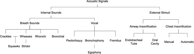
Recent developments in sensor technology and computational analysis methods enable new strategies to measure and interpret lung acoustic signals that originate internally, such as breathing or vocal sounds, or are externally introduced, such as in chest percussion or airway insonification. A better understanding of these sounds has resulted in a new instrumentation that allows for highly accurate as well as portable options for measurement in the hospital, in the clinic, and even at home. This review outlines the instrumentation for acoustic stimulation and measurement of the lungs. We first review the fundamentals of acoustic lung signals and the pathophysiology of the diseases that these signals are used to detect. Then, we focus on different methods of measuring and creating signals that have been used in recent research for pulmonary disease diagnosis. These new methods, combined with signal processing and modeling techniques, lead to a reduction in noise and allow improved feature extraction and signal classification. We conclude by presenting the results of human subject studies taking advantage of both the instrumentation and signal processing tools to accurately diagnose common lung diseases. This paper emphasizes the active areas of research within modern lung acoustics and encourages the standardization of future work in this field.
Figures
References
-
- Laennec RTH and Forbes J, A Treatise on the Diseases of the Chest, and on Mediate Auscultation. Samuel S. and William Wood, 1838.
-
- Cohen A, “Signal processing methods for upper airway and pulmonary dysfunction diagnosis,” IEEE Engineering in Medicine and Biology Magazine, vol. 9, no. 1, pp. 72–75, 1990. - PubMed
-
- Kompis M, Pasterkamp H, and Wodicka GR, “Acoustic imaging of the human chest,” Chest, vol. 120, no. 4, pp. 1309–1321, 2001. - PubMed
-
- Mor R, Kushnir I, Meyer J-J, Ekstein J, and Ben-Dov I, “Breath sound distribution images of patients with pneumonia and pleural effusion,” Respiratory care, vol. 52, no. 12, pp. 1753–1760, 2007. - PubMed
-
- Marshall A and Boussakta S, “Signal analysis of medical acoustic sounds with applications to chest medicine,” Journal of the Franklin Institute, vol. 344, no. 3–4, pp. 230–242, 2007.
Publication types
MeSH terms
Grants and funding
LinkOut - more resources
Full Text Sources
Other Literature Sources
Medical
