

Classification of Limbal Stem Cell Deficiency Using Clinical and Confocal Grading
- PMID: 30371569
- PMCID: PMC6279551
- DOI: 10.1097/ICO.0000000000001799
Classification of Limbal Stem Cell Deficiency Using Clinical and Confocal Grading
Abstract
Purpose: To grade the severity of limbal stem cell deficiency (LSCD) based on the extent of clinical presentation and central corneal basal epithelial cell density (BCD).
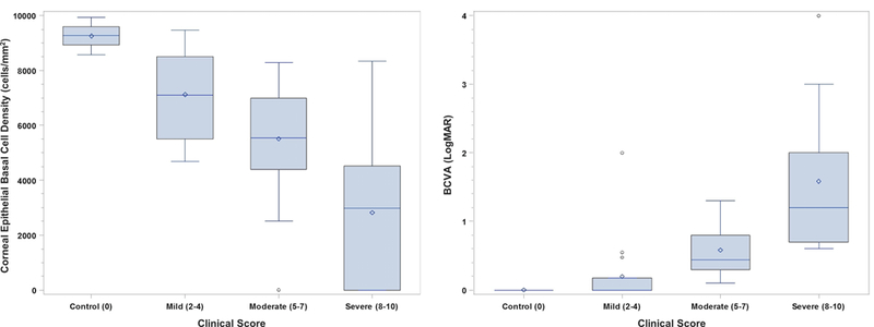
Methods: This is a retrospective observational comparative study of 48 eyes of 35 patients with LSCD and 9 eyes of 7 normal subjects (controls). Confocal images of the central cornea were acquired. A clinical scoring system was created based on the extent of limbal and corneal surface involvement. LSCD was graded as mild, moderate, and severe stages based on the clinical scores. The degree of BCD reduction was given a score of 0 to 3.
Results: Compared with BCD in controls, BCD decreased by 23.0%, 40.4%, and 69.5% in the mild, moderate, and severe stages of LSCD classified by the clinical scoring system, respectively. The degree of BCD reduction was positively correlated with larger limbal and corneal surface involvement and when the central visual axis was affected (all P ≤ 0.0005). Mean corrected distance visual acuity logarithm of the minimum angle of resolution was 0.0 ± 0.0 in control eyes, 0.2 ± 0.5 in mild LSCD, 0.6 ± 0.4 in moderate LSCD, and 1.6 ± 1.1 in severe LSCD (P < 0.0001). There was a significant correlation between a higher clinical score and corrected distance visual acuity logarithm of the minimum angle of resolution (rho = 0.82; P < 0.0001) and a greater decrease in BCD (rho = -0.78; P < 0.0001).
Conclusions: A clinical scoring system was developed to assess the extent of clinical presentation of LSCD. A classification system to grade the severity of LSCD can be established by combining the BCD score with the clinical score.
Conflict of interest statement
Figures


References
-
- Lavker RM, Tseng SC, Sun TT. Corneal epithelial stem cells at the limbus: looking at some old problems from a new angle. Exp Eye Res.2004;78:433–446. - PubMed
-
- Tseng SC. Concept and application of limbal stem cells. Eye (Lond).1989;3 ( Pt 2):141–157. - PubMed
-
- Dua HS, Azuara-Blanco A. Limbal stem cells of the corneal epithelium. Surv Ophthalmol.2000;44:415–425. - PubMed
Publication types
MeSH terms
Grants and funding
LinkOut - more resources
Full Text Sources
Other Literature Sources
Medical
