. 2018 Oct 1;59(12):5176-5187.
doi: 10.1167/iovs.18-25389.

Ocular Biometric Diurnal Rhythms in Emmetropic and Myopic Adults

Affiliations

Ocular Biometric Diurnal Rhythms in Emmetropic and Myopic Adults

Hannah J Burfield et al. Invest Ophthalmol Vis Sci. .

Abstract

Purpose: To investigate diurnal variations in anterior and posterior segment biometry and assess differences between emmetropic and myopic adults.

Methods: Healthy subjects (n = 42, 23-41 years old) underwent biometry and spectral-domain optical coherence tomography imaging (SD-OCT) every 4 hours for 24 hours. Subjects were in darkness from 11:00 PM to 7:00 AM. Central corneal thickness, corneal power, anterior chamber depth, lens thickness, vitreous chamber depth, and axial length were measured. Thicknesses of the total retina, photoreceptor outer segments + RPE, photoreceptor inner segments, and choroid over a 6-mm annulus were determined.

Results: All parameters except anterior chamber depth demonstrated significant diurnal variations, with no refractive error differences. Amplitude of choroid diurnal variation correlated with axial length (P = 0.05). Amplitude of axial length variation (35.71 ± 19.40 μm) was in antiphase to choroid variation (25.65 ± 2.01 μm, P < 0.001). The central 1-mm retina underwent variation of 5.03 ± 0.23 μm with a peak at 12 hours (P < 0.001), whereas photoreceptor outer segment + RPE thickness peaked at 4 hours and inner segment thickness peaked at 16 hours. Diurnal variations in retina and choroid were observed in the 3- and 6-mm annuli.

Conclusions: Diurnal rhythms in anterior and posterior segment biometry were observed over 24 hours in adults. Differences in baseline parameters were found between refractive error groups, and choroid diurnal variation correlated with axial length. The retina and choroid exhibited diurnal thickness variations in foveal and parafoveal regions.


Figures

Figure 1
Figure 1
(A) Radial line scan pattern used for SD-OCT imaging. (B) Choroid thickness map generated from semi-automated segmentation. (C) Segmentation lines included the inner limiting membrane (green), external limiting membrane (pink), inner segment/outer segment junction (yellow), Bruch's membrane (red), and choroid/sclera border (blue). (D) Data were binned by eccentricity into the central 1-mm region, 3-mm annulus, and 6-mm annulus.
Figure 2
Figure 2
Bland-Altman analysis for repeated measures of (A) total retina thickness and (B) choroid thickness for the central 1-mm region. Error bars represent 95% confidence interval of the limits of agreement.
Figure 3
Figure 3
(A) Total retina thickness for all subjects by eccentricity and quadrant (mean ± SE in μm). (B) Total retinal thickness across the horizontal meridian for emmetropic subjects (closed symbols) and myopic subjects (open symbols). (C) Choroid thickness for all subjects by eccentricity and quadrant (mean ± SE in μm). (D) Choroid thickness across the horizontal meridian for emmetropic subjects (closed symbols) and myopic subjects (open symbols).
Figure 4
Figure 4
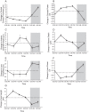
Diurnal changes from the mean (mean ± SE) over 24 hours for all subjects for (A) central corneal thickness (μm), (B) corneal power (D), (C) anterior chamber depth (μm), (D) vitreous chamber depth (μm), (E) lens thickness (μm), (F) calculated lens power (D), and (G) axial length (μm); shaded regions represent the dark period.
Figure 5
Figure 5
Diurnal changes from the mean (mean ± SE, μm) over 24 hours for all subjects for (A) total retina thickness in the central 1-mm diameter, (B) photoreceptor outer segment (OS) + RPE thickness in the central 1-mm diameter, (C) total retina thickness in the 3-mm annulus by quadrant, (D) photoreceptor inner retina thickness in the central 1-mm diameter, and (E) total retina thickness in the 6-mm annulus by quadrant; shaded regions represent the dark period.
Figure 6
Figure 6
Diurnal changes from the mean (mean ± SE, μm) over 24 hours for all subjects for choroid thickness in (A) the central 1-mm region, (B) the 3-mm annulus by quadrant, and (C) the 6-mm annulus by quadrant; shaded regions represent the dark period.
Figure 7
Figure 7
Diurnal changes from the mean (mean ± SE, μm) with fitted cosine functions over 24 hours for emmetropic subjects (closed symbols, solid lines) and myopic subjects (open symbols, dashed lines) for axial length (AL, black lines) and choroid thickness (square symbols, red lines); shaded region represents the dark period.

References

    1. Fujiwara T, Imamura Y, Margolis R, Slakter JS, Spaide RF. Enhanced depth imaging optical coherence tomography of the choroid in highly myopic eyes. Am J Ophthalmol. 2009;148:445–450. - PubMed
    1. Teberik K, Kaya M. Retinal and choroidal thickness in patients with high myopia without maculopathy. Pak J Med Sci. 2017;33:1438–1443. - PMC - PubMed
    1. Bhardwaj V, Rajeshbhai GP. Axial length, anterior chamber depth-a study in different age groups and refractive errors. J Clin Diagn Res. 2013;7:2211–2212. - PMC - PubMed
    1. Harper CL, Boulton ME, Bennett D, et al. Diurnal variations in human corneal thickness. Br J Ophthalmol. 1996;80:1068–1072. - PMC - PubMed
    1. Read SA, Collins MJ. Diurnal variation of corneal shape and thickness. Optom Vis Sci. 2009;86:170–180. - PubMed

Publication types