. 2018 Oct 29;19(1):783.
doi: 10.1186/s12864-018-5155-2.

Profiling the miRNA-mRNA-lncRNA interaction network in MSC osteoblast differentiation induced by (+)-cholesten-3-one

Affiliations

Profiling the miRNA-mRNA-lncRNA interaction network in MSC osteoblast differentiation induced by (+)-cholesten-3-one

Qiuke Hou et al. BMC Genomics. .

Abstract

Background: Our previous study showed that (+)-cholesten-3-one (CN) has the potential to induce the osteoblastic differentiation of mesenchymal stem cells (MSCs). However, the roles of CN in targeting miRNA-mRNA-lncRNA interactions to regulate osteoblast differentiation remain poorly understood.

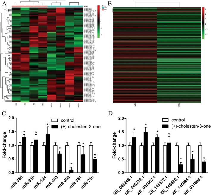
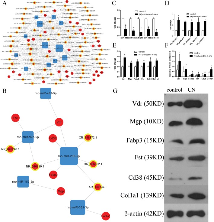
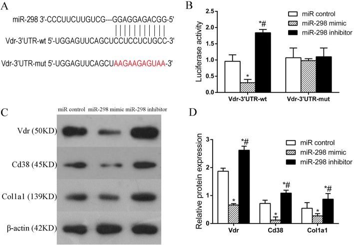
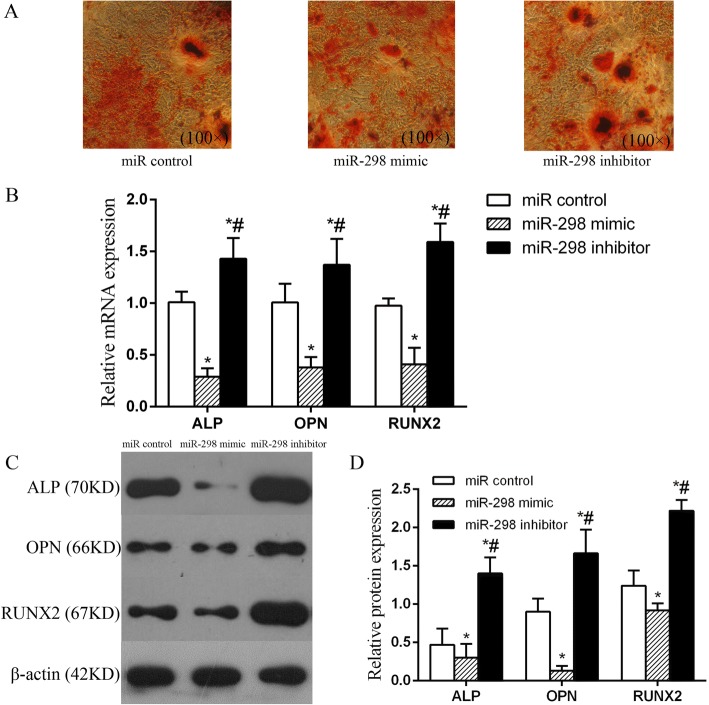
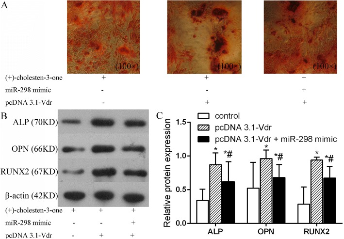
Results: A total of 77 miRNAs (36 upregulated and 41 downregulated) and 295 lncRNAs (281 upregulated and 14 downregulated) were significantly differentially expressed during CN-induced MSC osteogenic differentiation. Bioinformatic analysis identified that several pathways may play vital roles in MSC osteogenic differentiation, such as the vitamin D receptor signalling, TNF signalling, PI3K-Akt signalling, calcium signalling, and mineral absorption pathways. Further bioinformatic analysis revealed 16 core genes, including 6 mRNAs (Vdr, Mgp, Fabp3, Fst, Cd38, and Col1a1), 5 miRNAs (miR-483, miR-298, miR-361, miR-92b and miR-155) and 5 lncRNAs (NR_046246.1, NR_046239.1, XR_086062.1, XR_145872.1 and XR_146737.1), that may play important roles in regulating the CN-induced osteogenic differentiation of MSCs. Verified by the luciferase reporter, AR-S, qRT-PCR and western blot assays, we identified one miRNA (miR-298) that may enhance the osteogenic differentiation potential of MSCs via the vitamin D receptor signalling pathway.

Conclusions: This study revealed the global expression profile of miRNAs and lncRNAs involved in the Chinese medicine active ingredient CN-induced osteoblast differentiation of MSCs for the first time and provided a foundation for future investigations of miRNA-mRNA-lncRNA interaction networks to completely illuminate the regulatory role of CN in MSC osteoblast differentiation.

Keywords: (+)-cholesten-3-one; Mesenchymal stem cells; Osteoblastic differentiation; miRNA-mRNA-lncRNA.


Conflict of interest statement

Consent for publication

Not applicable.

Competing interests

The authors declare that they have no competing interests.

Publisher’s Note

Springer Nature remains neutral with regard to jurisdictional claims in published maps and institutional affiliations.

Figures

Fig. 1
Fig. 1
Heat map of miRNA and lncRNA differential expression during the CN-induced osteoblast differentiation of MSCs. a In total, 77 miRNAs were differentially expressed, including 36 that were upregulated and 41 that were downregulated. b In total, 295 lncRNAs were differentially expressed, including 281 that were upregulated and 14 that were downregulated. c qRT-PCR verification of the miRNA microarray results. Compared with the control group, the expression levels of miR-365, miR-330 and miR-124 were higher in CN-induced MSCs, and those of miR-483, miR-298, miR-361 and miR-296 were lower in CN-induced MSCs, which was consistent with the miRNA microarray results. d qRT-PCR verification of the lncRNA microarray results. Compared with the control group, the expression levels of NR_046246.1, NR_046239.1, XR_086062.1, and XR_145872.1 were higher in CN-induced MSCs, and those of NR_031896.1, XR_146480.1 and XR_145894.1 were lower in CN-induced MSCs, which was consistent with the lncRNA microarray results. P < 0.05, compared to the control group. The color scale shown on the top illustrates the relative expression level of the genes between two group: red denotes high expression levels, whereas green denotes low expression levels
Fig. 2
Fig. 2
Candidate mRNA predictions and CNC network construction during CN-induced MSC osteoblast differentiation. a In total, 443 mRNAs were predicted via mRBase targets based on the miRNA and lncRNA microarray. Seven mRNAs (Vdr, Mgp, Fst, Cd38, Nln, Rfc3 and Car9) were selected for validation via qRT-PCR, and the results were consistent with the predicted target genes. b CNC networks during the CN-induced osteoblast differentiation of MSCs. The dots represent mRNAs, the dots in circles represent lncRNAs, and the squares represent miRNAs. All the red dots represent upregulation, and all the blue dots represents downregulation. P < 0.05, compared to the control group
Fig. 3
Fig. 3
GO and KEGG analyses of predicted target genes during CN-induced MSC osteoblast differentiation. a GO analysis of 443 predicted target genes. In total, 174 biological processes were enriched; upregulation is shown in red, and downregulation is shown in blue. The y-axis shows the top ten biological processes, and the x-axis shows the negative logarithm of the p value (-LgP) of each biological process. b KEGG analysis of upregulated predicted target genes. c KEGG pathway analysis of downregulated predicted target genes. In both B & C, the y-axis shows the pathway category, and the x-axis shows the richness factor. A larger richness factor indicates greater enrichment. The size of the bubble indicates the number of genes in the pathway, and the colour of the bubble represents the range of P values
Fig. 4
Fig. 4
Core CNC networks during the CN-induced osteoblast differentiation of MSCs. a Core CNC network based on the 29 core genes obtained from GO cluster analysis and KEGG pathway analysis. b Sub-core CNC networks based on the 6 core genes obtained from online databases and core CNC networks. c qRT-PCR verification of miRNA expression. Compared with the control group, the expression levels of all the core miRNAs were lower in the CN-induced MSC group, which was consistent with the sub-core CNC network results. d qRT-PCR verification of lncRNA expression. Compared with the control group, the expression levels of all the core lncRNAs were higher in the CN-induced MSC group, which was consistent with the sub-core CNC network results. e qRT-PCR verification of mRNA expression. Compared with the control group, the expression levels of all the core mRNAs were higher in the CN-induced MSC group, which was consistent with the sub-core CNC network results. f and g Western blot verification of mRNA expression; the results were similar to the qRT-PCR results. P < 0.05, compared to the control group
Fig. 5
Fig. 5
miR-298 directly targets Vdr during CN-induced MSC osteoblast differentiation. a Predicted miR-298 target sequence in the 3’UTR of Vdr. b Luciferase reporter assay of MSCs co-transfected with wt or mut Vdr plasmids and miR control, miR-298 mimic or miR-298 inhibitor, which showed that overexpression of miR-298 remarkably inhibited the luciferase activity of the wild-type Vdr 3’UTR (Vdr-3’UTR-wt) construct but not that of the mutated Vdr 3’UTR (Vdr-3’UTR-mut) construct. c and d Western blot detection of Vdr, Cd38, and Col1a1 protein expression in MSCs transfected with miR-298 mimic or miR-298 inhibitor. miR control group: MSCs transfected with miR control; miR-298 mimic group: MSCs transfected with miR-298 mimic; miR-298 inhibitor group: MSCs transfected with miR-298 inhibitor. *P < 0.05 vs. the miR control group. #P < 0.05 vs. the miR-298 mimic group
Fig. 6
Fig. 6
miR-298 negatively regulated osteogenesis-related factors during CN-induced MSC osteoblast differentiation. a AR-S showed that more calcium mineral deposition was found in the miR-298 inhibitor group than in the miR control and miR-298 mimic groups; in other words, miR-298 suppressed MSC osteogenic differentiation. b qRT-PCR detection of the mRNA expression of osteogenic-related factors in MSCs transfected with miR-298 mimic or miR-298 inhibitor showing that the mRNA levels of the osteogenic-related factors were decreased in the miR-298 mimic group compared with those in the miR control group, whereas these levels were enhanced in the miR-298 inhibitor group. c and d Western blot detection of the protein expression of osteogenic-related factors in MSCs transfected with miR-298 mimic or miR-298 inhibitor; the results were similar to the qRT-PCR results. miR control group: MSCs transfected with miR control; miR-298 mimic group: MSCs transfected with miR-298 mimic; miR-298 inhibitor group: MSCs transfected with miR-298 inhibitor. *P < 0.05 vs. the miR control group. #P < 0.05 vs. the miR-298 mimic group
Fig. 7
Fig. 7
Vdr overexpression rescues the inhibitory effect of miR-298 during the CN-induced osteoblast differentiation of MSCs. a AR-S showed that more calcium mineral deposition was found in the CN + pcDNA 3.1-Vdr + miR-298 mimic group than in the CN group, while less calcium mineral deposition was found in the CN + pcDNA 3.1-Vdr group. Thus, pcDNA 3.1-Vdr rescued the inhibitory effect of miR-298 during the CN-induced osteoblast differentiation of MSCs. b & c Western blot detection of the protein expression of osteogenic-related factors in MSCs transfected with miR-298 mimic or pcDNA 3.1-Vdr; the results were similar to the AR-S results. Control group: MSCs transfected without pcDNA 3.1-Vdr or miR-298 mimic; pcDNA 3.1-Vdr group: MSCs transfected with pcDNA 3.1-Vdr; pcDNA 3.1-Vdr + miR-298 mimic group: MSCs co-transfected with pcDNA 3.1-Vdr and miR-298 mimic. *P < 0.05 vs. the CN group. #P < 0.05 vs. the miR-298 mimic group

Similar articles

Cited by

References

    1. Bianco P, Riminucci M, Gronthos S, Robey PG. Bone marrow stromal stem cells: nature, biology, and potential applications. Stem Cells. 2001;19(3):180–192. doi: 10.1634/stemcells.19-3-180. - DOI - PubMed
    1. Bianco P, Robey PG, Simmons PJ. Mesenchymal stem cells: revisiting history, concepts, and assays. Cell Stem Cell. 2008;2(4):313–319. doi: 10.1016/j.stem.2008.03.002. - DOI - PMC - PubMed
    1. Chen Q, Shou P, Zheng C, et al. Fate decision of mesenchymal stem cells: adipocytes or osteoblasts? Cell Death Differ. 2016;23(7):1128–1139. doi: 10.1038/cdd.2015.168. - DOI - PMC - PubMed
    1. Deng P, Zhou C, Alvarez R, Hong C, Wang CY. Inhibition of IKK/NF-kappaB signaling enhances differentiation of mesenchymal stromal cells from human embryonic stem cells. Stem Cell Rep. 2016;6(4):456–465. doi: 10.1016/j.stemcr.2016.02.006. - DOI - PMC - PubMed
    1. Hou QK, Huang YQ, Luo YW, et al. (+)-Cholesten-3-one induces osteogenic differentiation of bone marrow mesenchymal stem cells by activating vitamin D receptor. Exper Ther Med. 2017;13(5):1841–1849. doi: 10.3892/etm.2017.4200. - DOI - PMC - PubMed

MeSH terms