. 2018 Nov 13;115(46):E10849-E10858.
doi: 10.1073/pnas.1803377115. Epub 2018 Oct 29.

microRNA-378 promotes autophagy and inhibits apoptosis in skeletal muscle

Affiliations

microRNA-378 promotes autophagy and inhibits apoptosis in skeletal muscle

Yan Li et al. Proc Natl Acad Sci U S A. .

Abstract

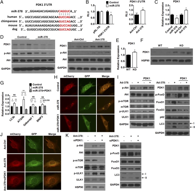
The metabolic regulation of cell death is sophisticated. A growing body of evidence suggests the existence of multiple metabolic checkpoints that dictate cell fate in response to metabolic fluctuations. However, whether microRNAs (miRNAs) are able to respond to metabolic stress, reset the threshold of cell death, and attempt to reestablish homeostasis is largely unknown. Here, we show that miR-378/378* KO mice cannot maintain normal muscle weight and have poor running performance, which is accompanied by impaired autophagy, accumulation of abnormal mitochondria, and excessive apoptosis in skeletal muscle, whereas miR-378 overexpression is able to enhance autophagy and repress apoptosis in skeletal muscle of mice. Our in vitro data show that metabolic stress-responsive miR-378 promotes autophagy and inhibits apoptosis in a cell-autonomous manner. Mechanistically, miR-378 promotes autophagy initiation through the mammalian target of rapamycin (mTOR)/unc-51-like autophagy activating kinase 1 (ULK1) pathway and sustains autophagy via Forkhead box class O (FoxO)-mediated transcriptional reinforcement by targeting phosphoinositide-dependent protein kinase 1 (PDK1). Meanwhile, miR-378 suppresses intrinsic apoptosis initiation directly through targeting an initiator caspase-Caspase 9. Thus, we propose that miR-378 is a critical component of metabolic checkpoints, which integrates metabolic information into an adaptive response to reduce the propensity of myocytes to undergo apoptosis by enhancing the autophagic process and blocking apoptotic initiation. Lastly, our data suggest that inflammation-induced down-regulation of miR-378 might contribute to the pathogenesis of muscle dystrophy.

Keywords: apoptosis; autophagy; miR-378; skeletal muscle.


Conflict of interest statement

The authors declare no conflict of interest.

Figures

Fig. 1.
Fig. 1.
miR-378/378* KO mice exhibit multiple abnormalities in skeletal muscle. (A) qPCR analysis of relative miR-378 levels in different tissues [brain (Bra), epididymal fat (Ep), heart (He), kidney (Kid), liver (Liv), lung (Lun), skeletal muscle (Mus), pancreas (Pan), spleen (Spl), and testis (Tes)] from C57BL/6J mice as indicated. (B) qPCR analysis of relative miR-378 levels in the TA muscle of fed and fasted mice. (C) Relative muscle weight (MW), including SOL, GAS, and TA weight, to body weight (BW) ratio of miR-378/378* KO and WT mice (n = 11–12). (D) Immunofluorescence staining of Caveolin (Left) and average muscle fiber area (Right) in the GAS muscle of KO and WT mice. (E) Maximal treadmill running distance for KO and WT mice (n = 5–6). (F) EM images of autophagosomes (arrowheads) and abnormal mitochondria in the GAS muscle of WT and KO mice, respectively. The areas indicated by boxes in Upper are shown at higher magnification in Lower. (Scale bars: F, Upper, 2 μm; F, Lower, 500 nm.) (G) qPCR analysis of the mRNA levels of LC3b, ATG12, ATG4b, Garbarapl 1, BNIP3, Beclin, and VPS34 in the GAS muscle of KO and WT mice. (H) Western blots of LC3 (LC3-I and LC3-II) as indicated by arrows in the GAS muscle of KO and WT mice. (I) Western blots of LC3 (LC3-I and LC3-II) as indicated by arrows in the GAS muscle of fed or fasted KO and WT mice. (J) Western blots of PARP in the GAS muscle of KO and WT mice. Cleaved PARP proteins are indicated by an arrow. Means ± SEM are shown. *P < 0.05; **P < 0.01; ***P < 0.001.
Fig. 2.
Fig. 2.
miR-378 promotes autophagy in muscle cells. (A) qPCR analysis of relative miR-378 levels in C2C12 myotubes cultured in (Left) No Glu or (Right) KRB for indicated time periods. (B) Western blots of LC3 (LC3-I and LC3-II) as indicated by arrows in Ad-378–infected C2C12 myotubes cultured in (Left) No Glu or (Right) KRB as indicated. (C) Western blots of LC3 (LC3-I and LC3-II) as indicated by arrows in C2C12 myotubes transfected with miR-378 mimics (miR-378) and control oligos (Control) or Ant-378 and antagomir control (Ant-Ctrl) as indicated. (D) Immunofluorescence staining of LC3 (red) in C2C12 myoblasts transfected with miR-378 mimics (miR-378), control oligos (Control), Ant-378, or Ant-Ctrl as indicated. (E) Immunofluorescence images of LC3 in Ad-mCherry-GFP-LC3–infected C2C12 myoblasts transfected with miR-378 mimics (miR-378) or control oligos (Control; Left) and Ant-378 or Ant-Ctrl (Right) as indicated. (F) Western blots of p62 in C2C12 myotubes transfected with miR-378 mimics (miR-378), control oligos (Control), Ant-378, or Ant-Ctrl as indicated. (G) Western blots of p-mTOR, mTOR, p-ULK1 (Ser757), ULK1, p-FoxO [p-FoxO1 (Thr24) and p-FoxO3 (Thr32)], FoxO1, and FoxO3 in C2C12 myotubes transfected with miR-378 mimics (miR-378), control oligos (Control), Ant-378, or Ant-Ctrl as indicated. (H) Western blots of LC3 (LC3-I and LC3-II) as indicated by arrows in Ad-378–infected C2C12 myotubes in the presence or absence of Baf A1 (Upper) or CQ (Lower) as indicated. Means ± SEM are shown. **P < 0.01; ***P < 0.001.
Fig. 3.
Fig. 3.
PDK1 mediates the effect of miR-378 on autophagy in skeletal muscle. (A) Sequence alignment of miR-378 and the PDK1-3′UTR from various species. (B) Luciferase assay showing the effect of miR-378 mimics (miR-378) transfection (Left) or Ant-378 transfection (Right) on the activity of the reporter containing the PDK1-3′UTR with a putative miR-378 recognition element in C2C12 myoblasts. (C and D) qPCR analysis of relative PDK1 mRNA levels (C) and Western blots (D) of PDK1 and p-Akt (Thr-308) in C2C12 myotubes transfected with miR-378 mimics (miR-378), control oligos (Control), Ant-378, or antagomir control (Ant-Ctrl) as indicated. (E and F) qPCR analysis of relative PDK1 mRNA levels (E) and Western blots of PDK1 (F) in the GAS muscle of KO and WT mice. (G) qPCR analysis of the mRNA levels of LC3b, ATG4b, Gabarapl 1, and BNIP3 in C2C12 myotubes transfected with miR-378 mimics (miR-378) or control oligos (Control) with or without PDK1 expression plasmids as indicated. (H) Immunofluorescence images of LC3 in Ad-mCherry-GFP-LC3–infected C2C12 myoblasts transfected with control oligos (Control), miR-378 mimics (miR-378) alone, or miR-378 mimics together with PDK1 expression plasmids. (I) Western blots of PDK1, p-Akt (Thr-308), Akt, p-mTOR, mTOR, p-ULK1 (Ser757), ULK1, p-FoxO [p-FoxO1 (Thr24) and p-FoxO3 (Thr32)], FoxO1, FoxO3, p62, and LC3 in Ad-378–infected C2C12 myotubes transfected with or without PDK1 expression plasmids as indicated. (J) Immunofluorescence images of LC3 in Ad-mCherry-GFP-LC3–infected C2C12 myoblasts transfected with Ant-Ctrl, Ant-378 alone, or Ant-378 together with siRNA specific for PDK1 (siPDK1). (K) Western blots of PDK1, p-Akt (Thr-308), Akt, p-mTOR, mTOR, p-ULK1 (Ser757), ULK1, p-FoxO [p-FoxO1 (Thr24) and p-FoxO3 (Thr32)], FoxO1, FoxO3, and LC3 in C2C12 myotubes transfected with Ant-378 and/or siPDK1 as indicated. Means ± SEM are shown. NS, not significant; RLU, relative luciferase units. *P < 0.05; **P < 0.01; ***P < 0.001.
Fig. 4.
Fig. 4.
CASP9 is a direct target gene of miR-378 in skeletal muscle. (A) Sequence alignment of miR-378 and the CASP9-3′UTR from various species. (B) Luciferase assay showing the effect of miR-378 mimics (miR-378) transfection (Left) or Ant-378 transfection (Right) on the activity of CASP9-3′UTR with the putative miR-378 binding site in C2C12 myoblasts. RLU, relative luciferase units. (C and D) qPCR analysis of relative CASP9 mRNA levels (C) and Western blots (D) of CASP9 in C2C12 myotubes transfected with miR-378 mimics (miR-378), control oligos (Control), Ant-378, or antagomir control (Ant-Ctrl) as indicated. (E) Immunofluorescence staining of cleaved CASP3 (red) in C2C12 myoblasts transfected with miR-378 mimics (miR-378), control oligos (Control), Ant-378, or Ant-Ctrl as indicated. (F) Western blots of PARP in C2C12 myotubes transfected with miR-378 mimics (miR-378), control oligos (Control), Ant-378, or Ant-Ctrl as indicated. (GI) qPCR analysis of relative CASP9 mRNA levels (G), Western blots of CASP9 and cleaved CASP9 (H), and relative CASP9 activity (I) in the GAS muscle of KO and WT mice. (J) Western blots of CASP3 and cleaved CASP3 in the GAS muscle of KO and WT mice. (K) Maximal treadmill running distance for KO mice treated with or without z-VAD (n = 6). (L) Western blots of p62, LC3, PARP, CASP3, and cleaved CASP3 in the GAS muscle of KO mice treated with or without z-VAD as indicated. LC3-I, LC3-II, and cleaved PARP proteins are indicated by arrows. Means ± SEM are shown. *P < 0.05; **P < 0.01.
Fig. 5.
Fig. 5.
Overexpression of miR-378 induces autophagy and represses apoptosis in skeletal muscle. (A) qPCR analysis of relative miR-378 levels in the GAS and TA muscles of mice treated with agomir-378 or control. (B) qPCR analysis of relative mRNA levels of PDK1 and CASP9 in the GAS muscle of mice treated with agomir-378 or control. (CE) Western blots of CASP9, cleaved CASP9, PDK1, and LC3 (C); CASP3 and cleaved CASP3 (D); and PARP (E) in the GAS muscle of mice treated with agomir-378 or control. LC3-I, LC3-II, and cleaved PARP proteins are indicated by arrows. (F) qPCR analysis of LC3b, ATG12, BNIP3, BNIP3l, Cathepsin L, Atrogin 1, VPS34, and Beclin in the GAS muscle of mice treated with agomir-378 or control. (G) Western blots of p62 and LC3 (LC3-I and LC3-II) in the GAS muscle of fed or fasted mice treated with agomir-378 or control as indicated. Means ± SEM are shown. *P < 0.05; **P < 0.01.
Fig. 6.
Fig. 6.
Schematic diagram of the working model of miR-378 in the coordination of autophagy and apoptosis in skeletal muscle. mTORC1, mammalian target of rapamycin complex 1.

References

    1. Green DR, Galluzzi L, Kroemer G. Cell biology. Metabolic control of cell death. Science. 2014;345:1250256. - PMC - PubMed
    1. Mariño G, Niso-Santano M, Baehrecke EH, Kroemer G. Self-consumption: The interplay of autophagy and apoptosis. Nat Rev Mol Cell Biol. 2014;15:81–94. - PMC - PubMed
    1. Wirawan E, Vanden Berghe T, Lippens S, Agostinis P, Vandenabeele P. Autophagy: For better or for worse. Cell Res. 2012;22:43–61. - PMC - PubMed
    1. Levine B, Kroemer G. Autophagy in the pathogenesis of disease. Cell. 2008;132:27–42. - PMC - PubMed
    1. Roos WP, Thomas AD, Kaina B. DNA damage and the balance between survival and death in cancer biology. Nat Rev Cancer. 2016;16:20–33. - PubMed

Publication types

MeSH terms