. 2018 Nov 13;115(46):11796-11801.
doi: 10.1073/pnas.1813143115. Epub 2018 Oct 29.

Engineering yeast endosymbionts as a step toward the evolution of mitochondria

Affiliations

Engineering yeast endosymbionts as a step toward the evolution of mitochondria

Angad P Mehta et al. Proc Natl Acad Sci U S A. .

Abstract

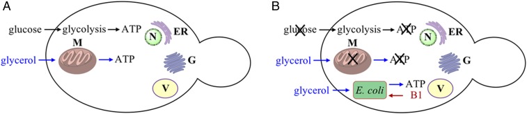
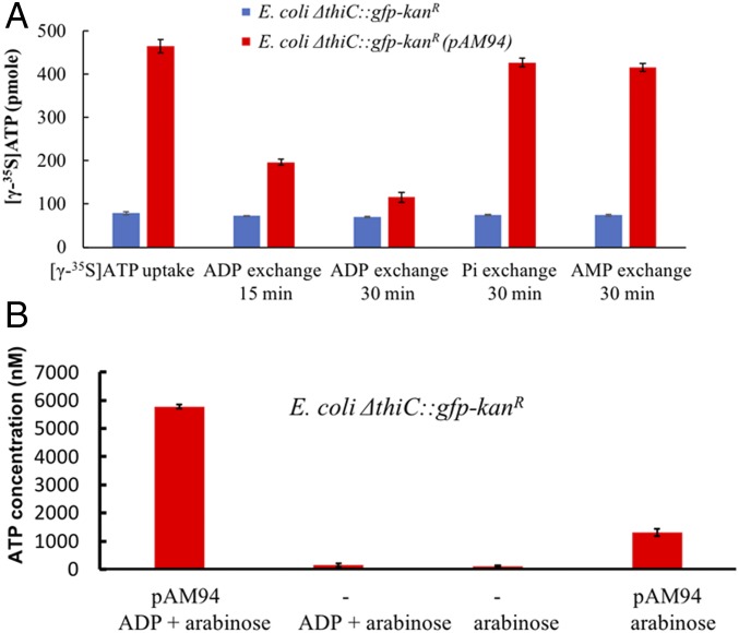
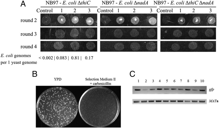
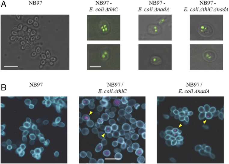
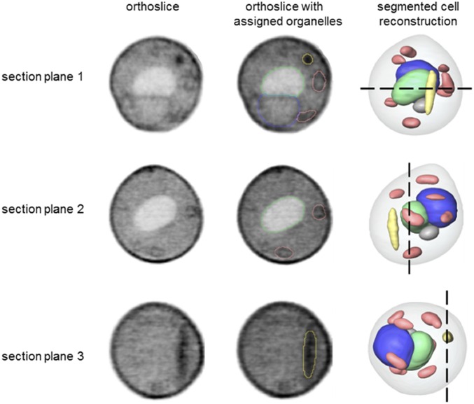
It has been hypothesized that mitochondria evolved from a bacterial ancestor that initially became established in an archaeal host cell as an endosymbiont. Here we model this first stage of mitochondrial evolution by engineering endosymbiosis between Escherichia coli and Saccharomyces cerevisiae An ADP/ATP translocase-expressing E. coli provided ATP to a respiration-deficient cox2 yeast mutant and enabled growth of a yeast-E. coli chimera on a nonfermentable carbon source. In a reciprocal fashion, yeast provided thiamin to an endosymbiotic E. coli thiamin auxotroph. Expression of several SNARE-like proteins in E. coli was also required, likely to block lysosomal degradation of intracellular bacteria. This chimeric system was stable for more than 40 doublings, and GFP-expressing E. coli endosymbionts could be observed in the yeast by fluorescence microscopy and X-ray tomography. This readily manipulated system should allow experimental delineation of host-endosymbiont adaptations that occurred during evolution of the current, highly reduced mitochondrial genome.

Keywords: ADP/ATP translocase; endosymbiotic theory; evolution; mitochondria.


Conflict of interest statement

The authors declare no conflict of interest.

Figures

Fig. 1.
Fig. 1.
Strategy to engineer S. cerevisiae–E. coli endosymbiont chimera. (A) Wild-type S. cerevisiae can grow on medium with glucose or glycerol due to ATP production by glycolysis in the cytoplasm and oxidative phosphorylation in mitochondria. (B) Yeast cells with a defect in oxidative phosphorylation cannot utilize glycerol for ATP synthesis and cannot grow in the absence of glucose. Introduction of E. coli-expressing ADP/ATP translocase and SNARE proteins into such mutant yeast can restore yeast growth with glycerol as the sole carbon source. Growth of intracellular E. coli is dependent on thiamin diphosphate (vitamin B1) provided by yeast. ER, endoplasmic reticulum; G, Golgi apparatus; M, mitochondria; N, nucleus; V, vacuole.
Fig. 2.
Fig. 2.
Release of ATP by E. coli cells encoding ADP/ATP translocase. (A) Cellular [γ-35S]ATP uptake/release by E. coli cells expressing the UWE25 ADP/ATP translocase (pAM94 plasmid) in the presence of 1 mM arabinose. Cellular [γ-35S]ATP was released when E. coli cells expressing the ADP/ATP translocase were challenged with extracellular ADP (10 mM), but not with phosphate (Pi) or AMP (each at 10 mM). (B) Release of ATP into the growth medium by E. coli cells expressing the UWE25 ADP/ATP translocase (pAM94 plasmid) in presence of 20 µM ADP and 1 mM arabinose. The ATP concentration in the medium was determined by luciferase assay. Data bars show a mean of three technical replicates; error bars represent SE of the mean.
Fig. 3.
Fig. 3.
S. cerevisiaeE. coli chimeras have a partially rescued respiration-competent phenotype. (A) Growth of S. cerevisiae cox2-60E. coli chimeras on medium containing glycerol as the sole carbon source, selection medium III. No growth was observed for parent cox2-60 yeast lacking intracellular E. coli (control). Three different chimera colonies growing during successive rounds of plating are shown for each S. cerevisiaeE. coli chimera. Number of E. coli genomes per one yeast genome was determined by qPCR for E. coli ΔthiC chimeras from the fourth round of growth. (B) A single cell suspension of S. cerevisiae cox2-60E. coli nadA chimera culture formed a comparable number of colonies on nonselective (YPD) and selective medium (selection medium II) plates. (C) Total DNAs isolated from colonies grown on selection medium II in B contain E. coli-encoded gfp gene. Ten random colonies (labeled 1–10) were PCR amplified for presence of gfp and MATa genes.
Fig. 4.
Fig. 4.
Imaging intracellular endosymbiont E. coli by fluorescent microscopy. (A) TIRF microscopic images of chimeric cells (Right) and control yeast cells (Left). Two representative cells of indicated chimera type are shown. All panels are merged images of TIRF (green) and differential interference contrast (grayscale). (Scale bar in the NB97 panel, 10 µm; scale bar in the NB97–E. coli ΔthiC panel, 5 µm.) (B) Confocal fluorescence microscopy images of control and chimeric yeast–E. coli cells. Yeast cell wall was stained with Con A-FITC (blue) and bacterial rRNA with EUB338-Cy3 probe (purple). Yellow arrowheads indicate examples of EUB338-positive yeast cells. (Scale bar in the Middle, 10 µm.)
Fig. 5.
Fig. 5.
Ultrastructural features of S. cerevisiaeE. coli ΔnadA chimera cells. Segmented reconstruction of a S. cerevisiaeE. coli ΔnadA cell viewed from three different perspectives (section planes 1–3). Orthoslices (Left) at positions indicated by the black dashed lines in a reconstructed cell (Right) are shown for each perspective. The same orthoslices are overlaid in the Center column with outlines indicating segmented organelle assignment. The gray values were generated using LACs, with black corresponding to the highest LAC value. Organelle color key: green, vacuole; blue, nucleus; salmon, mitochondria; and yellow, high LAC value, bacteria-like structure that remained unassigned after segmentation.

References

    1. Margulis L. Origin of Eukaryotic Cells: Evidence and Research Implications for a Theory of the Origin and Evolution of Microbial, Plant and Animal Cells on the Precambrian Earth. Yale Univ Press; New Haven, CT: 1970.
    1. Kutschera U, Niklas KJ. The modern theory of biological evolution: An expanded synthesis. Naturwissenschaften. 2004;91:255–276. - PubMed
    1. Gray MW. Mitochondrial evolution. Cold Spring Harb Perspect Biol. 2012;4:a011403. - PMC - PubMed
    1. Archibald JM. Endosymbiosis and eukaryotic cell evolution. Curr Biol. 2015;25:R911–R921. - PubMed
    1. Javaux EJ, Knoll AH, Walter MR. Morphological and ecological complexity in early eukaryotic ecosystems. Nature. 2001;412:66–69. - PubMed

Publication types

MeSH terms