

Microtubule-severing enzymes: From cellular functions to molecular mechanism
- PMID: 30373906
- PMCID: PMC6279391
- DOI: 10.1083/jcb.201612104
Microtubule-severing enzymes: From cellular functions to molecular mechanism
Abstract
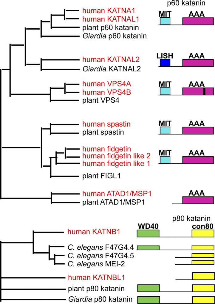
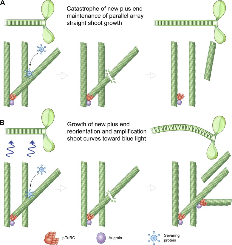
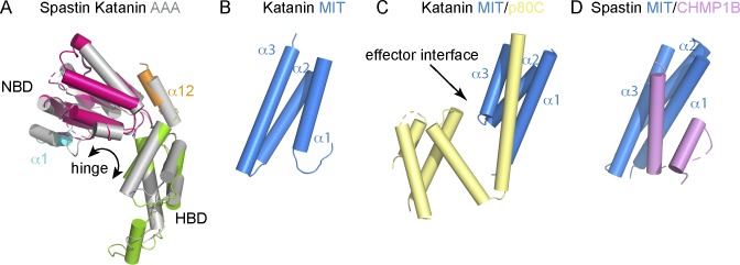
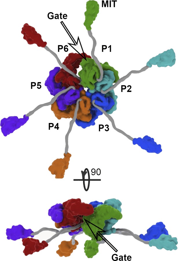
Microtubule-severing enzymes generate internal breaks in microtubules. They are conserved in eukaryotes from ciliates to mammals, and their function is important in diverse cellular processes ranging from cilia biogenesis to cell division, phototropism, and neurogenesis. Their mutation leads to neurodegenerative and neurodevelopmental disorders in humans. All three known microtubule-severing enzymes, katanin, spastin, and fidgetin, are members of the meiotic subfamily of AAA ATPases that also includes VPS4, which disassembles ESCRTIII polymers. Despite their conservation and importance to cell physiology, the cellular and molecular mechanisms of action of microtubule-severing enzymes are not well understood. Here we review a subset of cellular processes that require microtubule-severing enzymes as well as recent advances in understanding their structure, biophysical mechanism, and regulation.
© 2018 McNally and Roll-Mecak.
Figures
Similar articles
-
[Microtubule severing proteins - structure and activity regulation].Postepy Biochem. 2016;62(1):46-51. Postepy Biochem. 2016. PMID: 28132444 Review. Polish.
-
[Role of microtubule severing proteins in cytoskeleton reorganization].Postepy Biochem. 2016;62(1):52-59. Postepy Biochem. 2016. PMID: 28132445 Review. Polish.
-
In Vitro Reconstitution Assays of Microtubule Amplification and Lattice Repair by the Microtubule-Severing Enzymes Katanin and Spastin.Methods Mol Biol. 2020;2101:27-38. doi: 10.1007/978-1-0716-0219-5_3. Methods Mol Biol. 2020. PMID: 31879896 Free PMC article.
-
Fidgetin binds spastin to attenuate the microtubule-severing activity.Biochim Biophys Acta Mol Cell Res. 2025 Feb;1872(2):119890. doi: 10.1016/j.bbamcr.2024.119890. Epub 2024 Dec 15. Biochim Biophys Acta Mol Cell Res. 2025. PMID: 39681249
-
Purification and biophysical analysis of microtubule-severing enzymes in vitro.Methods Cell Biol. 2013;115:191-213. doi: 10.1016/B978-0-12-407757-7.00013-X. Methods Cell Biol. 2013. PMID: 23973074
Cited by
-
SUMOylation of nuclear receptor Nor1/NR4A3 coordinates microtubule cytoskeletal dynamics and stability in neuronal cells.Cell Biosci. 2024 Jul 13;14(1):91. doi: 10.1186/s13578-024-01273-x. Cell Biosci. 2024. PMID: 38997783 Free PMC article.
-
The model of local axon homeostasis - explaining the role and regulation of microtubule bundles in axon maintenance and pathology.Neural Dev. 2019 Nov 9;14(1):11. doi: 10.1186/s13064-019-0134-0. Neural Dev. 2019. PMID: 31706327 Free PMC article. Review.
-
Mouse KL2 is a unique MTSE involved in chromosome-based spindle organization and regulated by multiple kinases during female meiosis.J Biomed Res. 2024 May 29;38(5):485-499. doi: 10.7555/JBR.37.20230290. J Biomed Res. 2024. PMID: 38808565 Free PMC article.
-
KATANIN-mediated microtubule severing is required for MTOC organisation and function in Marchantia polymorpha.Development. 2024 Oct 15;151(20):dev202672. doi: 10.1242/dev.202672. Epub 2024 Apr 30. Development. 2024. PMID: 38572965 Free PMC article.
-
Epsilon tubulin is an essential determinant of microtubule-based structures in male germ cells.EMBO Rep. 2024 Jun;25(6):2722-2742. doi: 10.1038/s44319-024-00159-w. Epub 2024 May 21. EMBO Rep. 2024. PMID: 38773322 Free PMC article.
References
Publication types
MeSH terms
Substances
Associated data
- Actions
- Actions
- Actions
- Actions
- Actions
- Actions
- Actions
