

Calcineurin promotes APC/C activation at meiotic exit by acting on both XErp1 and Cdc20
- PMID: 30373936
- PMCID: PMC6280790
- DOI: 10.15252/embr.201846433
Calcineurin promotes APC/C activation at meiotic exit by acting on both XErp1 and Cdc20
Abstract
Vertebrate oocytes await fertilization arrested at metaphase of the second meiotic division. Fertilization triggers a transient calcium wave, which induces the activation of the anaphase-promoting complex/cyclosome (APC/C) and its co-activator Cdc20 resulting in the destruction of cyclin B and hence meiotic exit. Two calcium-dependent enzymes are implicated in fertilization-induced APC/CCdc20 activation: calcium-/calmodulin-dependent kinase type II (CaMKII) and calcineurin (CaN). While the role of CaMKII in targeting the APC/C inhibitor XErp1/Emi2 for destruction is well-established, it remained elusive how CaN affects APC/CCdc20 activation. Here, we discover that CaN contributes to APC/CCdc20 activation in Xenopus laevis oocytes by two independent but interrelated mechanisms. First, it facilitates the degradation of XErp1 by dephosphorylating it at a site that is part of a phosphorylation-dependent recruiting motif for PP2A-B'56, which antagonizes inhibitory phosphorylation of XErp1. Second, it dephosphorylates Cdc20 at an inhibitory site, thereby supporting its APC/C-activating function. Thus, our comprehensive analysis reveals that CaN contributes to timely APC/C activation at fertilization by both negatively regulating the APC/C inhibitory activity of XErp1 and positively regulating the APC/C-activating function of Cdc20.
Keywords: Xenopus laevis; APC/C; Calcineurin; Cdc20; Meiosis; XErp1.
© 2018 The Authors.
Figures
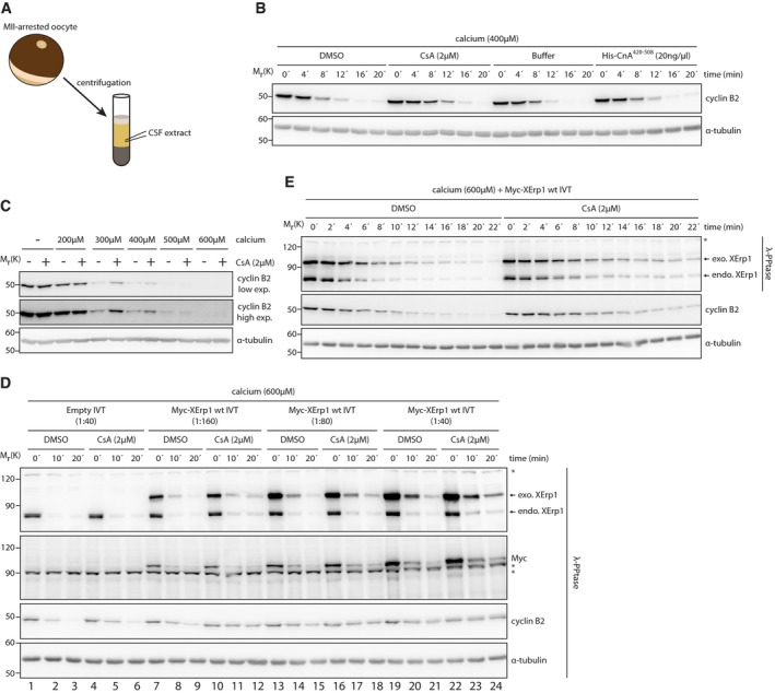
Scheme for the preparation of CSF extract.
CSF extract was treated with DMSO, CsA, buffer or His‐CnA420–508 at the indicated concentrations. Meiotic exit was induced by calcium addition, and samples were taken at the indicated time points. Samples were immunoblotted for cyclin B2. The cyclin B2 membrane was stripped and reprobed for α‐tubulin.
CSF extract was treated with DMSO or CsA. Both reactions were divided and supplemented with the indicated amounts of calcium or H2O. After 20 min, samples were taken and immunoblotted for cyclin B2 for which a low and high exposure is shown. The cyclin B2 membrane was stripped and reprobed for α‐tubulin.
CSF extract was supplemented with Myc‐XErp1 wt IVT at the indicated dilutions. An empty IVT reaction not expressing Myc‐XErp1 served as control. All reactions were divided and treated with DMSO or CsA. Calcium was added to all reactions, and samples were taken at the indicated time points, treated with λ‐phosphatase and immunoblotted for XErp1, the Myc‐tag, cyclin B2, and α‐tubulin. Asterisks indicate unspecific bands.
CSF extract was supplemented with Myc‐XErp1 wt IVT. The reaction was divided and treated with DMSO or CsA. Both reactions were treated with calcium, and samples were taken at the indicated time points and as indicated incubated with λ‐phosphatase. Samples were immunoblotted for XErp1 and cyclin B2. The cyclin B2 membrane was stripped and reprobed for α‐tubulin. Asterisk indicates unspecific bands.
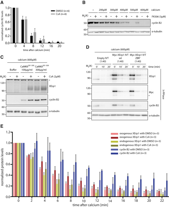
Quantification of cyclin B2 degradation in DMSO‐ or CsA‐treated CSF extracts after calcium addition as shown in Fig 1B. Values were normalized to t = 0 min and are given as mean ± standard deviation of six independent biological replicates. The P‐value for the 8 min timepoint (DMSO/CsA) is 0.08 (unpaired two‐sided t‐test with unequal variance).
CSF extract was treated with DMSO or FK506. Both reactions were divided and supplemented with the indicated amounts of calcium or H2O. After 20 min, samples were taken and immunoblotted for cyclin B2. The cyclin B2 membrane was stripped and reprobed for α‐tubulin.
CSF extract was treated with control buffer or the indicated amounts of CaM Kinase II Inhibitor 281–309 (CaMKII281–309). Extracts were divided and treated with DMSO or CsA before addition of calcium. After 15 min, samples were taken and immunoblotted for XErp1 and cyclin B2. The cyclin B2 membrane was stripped and reprobed for α‐tubulin. Asterisk indicates unspecific bands.
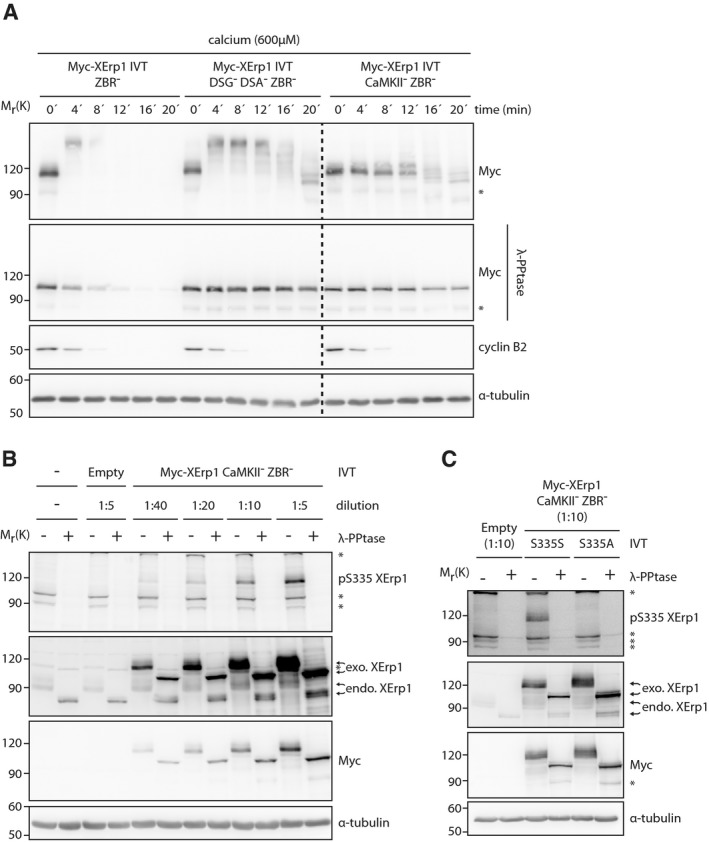
CSF extract was treated with Myc‐XErp1 IVT that was either wild‐type or mutated to alanine at C583 (ZBR−). An empty IVT not expressing XErp1 was added as control. Meiotic exit was induced by addition of calcium, and samples were taken at the indicated time points, treated with λ‐phosphatase and immunoblotted for XErp1, the Myc‐tag, cyclin B2, and α‐tubulin. Asterisk indicates unspecific bands.
Quantification of exogenous XErp1, endogenous XErp1, and cyclin B2 in DMSO‐ or CsA‐treated CSF extracts after calcium addition as shown in Fig 1E. Values were normalized to t = 0 min and are given as mean ± standard deviation of three independent biological replicates. The P‐values for the 8 min timepoint are 0.07 (exo. XErp1, DMSO/CsA), 0.26 (endo. XErp1, DMSO/CsA), and 0.17 (cyclin B2, DMSO/CsA) using an unpaired two‐sided t‐test with unequal variance.
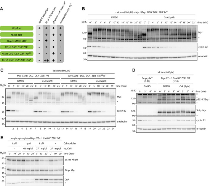
Schematic representation of the XErp1 variants and their characteristics used in this study.
CSF extract was supplemented with Myc‐XErp1 DSG− DSA− ZBR− (S33N S38N S284N S288N C583A) IVT and as indicated treated with DMSO or CsA. Both reactions were treated with calcium and samples were taken at the indicated time points. Samples were immunoblotted for the Myc‐tag, cyclin B2, and α‐tubulin. A pseudocolored representation of the Myc immunoblot is shown in Fig EV2A. Arrow marks the meiotic phosphorylation state of Myc‐XErp1. Asterisk indicates unspecific bands.
CSF extract was supplemented with Myc‐XErp1 DSG− DSA− ZBR− (S33N S38N S284N S288N C583A) IVT that was either wild‐type or mutated to alanine at the p90RSK‐target sites S335, T336, and S342 (Rsk3A). Both reactions were divided and treated with DMSO or CsA. Calcium was added, and samples were taken at the indicated time points. Samples were immunoblotted for the Myc‐tag and cyclin B2. The cyclin B2 membrane was stripped and reprobed for α‐tubulin. Asterisk indicates unspecific bands.
CSF extract was treated with Myc‐XErp1 CaMKII− ZBR− (T195A C583A) IVT. An empty IVT reaction not expressing XErp1 was used as control. The extracts were treated with DMSO or CsA as indicated. Calcium was added, and samples were taken at the indicated time points and immunoblotted for cyclin B2, pSer335 XErp1, and α‐tubulin. The pSer335 XErp1 membrane was stripped and reprobed for the Myc‐tag. Asterisks indicate unspecific bands.
Myc‐XErp1 CaMKII− ZBR− (T195A C583A) IVT was in vitro phosphorylated by recombinant PKA and isolated by α‐Myc immunoprecipitation. The immunoprecipitate was supplemented with calcium and treated with recombinant hs_CaN and calmodulin as indicated. Samples were taken at the indicated time points and immunoblotted for the catalytic calcineurin subunit CnA and pSer335 XErp1. The pSer335 XErp1 membrane was stripped and reprobed for the Myc‐tag. Asterisk indicates the IgG heavy chain.
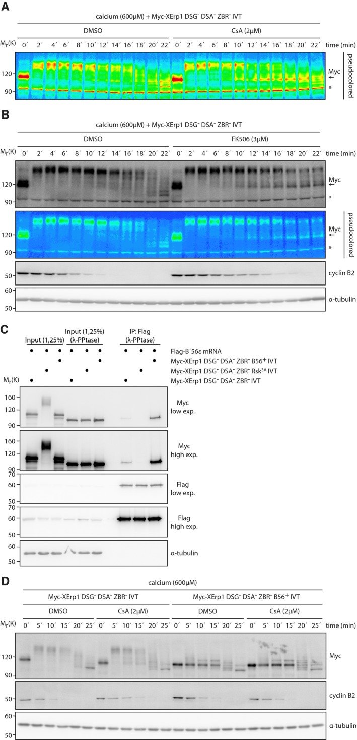
Pseudocolor representation of the Myc immunoblot shown in Fig 2B. Asterisk indicates unspecific bands.
CSF extract was supplemented with Myc‐XErp1 DSG− DSA− ZBR− (S33N S38N S284N S288N C583A) IVT. The reaction was divided and treated with DMSO or FK506. Both reactions were treated with calcium, and samples were taken at the indicated time points. Samples were immunoblotted for the Myc‐tag, cyclin B2, and α‐tubulin. A pseudocolored representation of the Myc immunoblot is shown. Arrow marks the meiotic phosphorylation state of Myc‐XErp1. Asterisk indicates unspecific bands.
CSF extract was treated with mRNA encoding Flag ‐B'56ε. The extract was divided and treated with Myc‐XErp1 IVT carrying the indicated combinations of the mutations DSG− DSA− ZBR− (S33N S38N S284N S288N C583A), Rsk3A (S335A T336A S342A), and B56+ (S335P L337I R340E G341E S342E). Flag‐B'56ε was immunoprecipitated, and the input and pellet (IP: Flag) samples were treated with λ‐phosphatase as indicated. The samples were immunoblotted for the Myc‐tag, the Flag‐tag, and α‐tubulin. Low and high exposures are shown for the Myc‐tag and the Flag‐tag.
CSF extract was supplemented with Myc‐XErp1 IVT carrying the indicated combinations of the mutations DSG− DSA− ZBR− (S33N S38N S284N S288N C583A) and B56+ (S335P L337I R340E G341E S342E). Both reactions were divided and treated with DMSO or CsA. Calcium was added, and samples were taken at the indicated time points. Samples were immunoblotted for the Myc‐tag, cyclin B2, and α‐tubulin.
CSF extract was treated with Myc‐XErp1 IVT carrying the indicated combinations of the mutations DSG− (S33N S38N), DSA− (S284N S288N), ZBR− (C583A) and CaMKII− (T195A). Calcium was added, samples were taken at the indicated time points and as indicated treated with λ‐phosphatase. Samples were immunoblotted for the Myc‐tag and cyclin B2. The cyclin B2 membrane was stripped and reprobed for α‐tubulin. Asterisk indicates unspecific bands. Several lanes were removed at the dashed line.
CSF extract was treated with Myc‐XErp1 CaMKII− ZBR− (T195A C583A) IVT at the indicated dilutions. An empty IVT not expressing XErp1 and an untreated condition were used as controls. Samples were taken, treated as indicated with λ‐phosphatase and immunoblotted for XErp1, pSer335 XErp1, and α‐tubulin. The XErp1 membrane was stripped and reprobed for the Myc‐tag. Asterisks indicate unspecific bands.
CSF extract was treated with Myc‐XErp1 CaMKII− ZBR− (T195A C583A) IVT that was either wild‐type or mutated to alanine at Ser335. An empty IVT reaction not expressing Myc‐XErp1 served as control. Samples were taken, treated as indicated with λ‐phosphatase and immunoblotted for XErp1, the Myc‐tag, pSer335 XErp1, and α‐tubulin. Asterisks indicate unspecific bands.
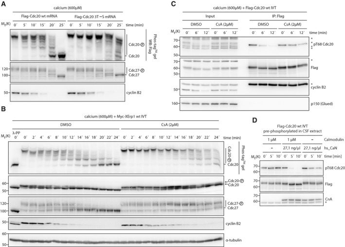
CSF extract was supplemented with mRNA encoding Flag‐Cdc20 that was either wild‐type or mutated to serine at Thr64, Thr68, and Thr79 (3T→S). After expression, calcium was added, samples were taken at the indicated time points and resolved by either conventional or Phos‐tag™ SDS–PAGE. Samples were immunoblotted for the Flag‐tag, Cdc27, and cyclin B2.
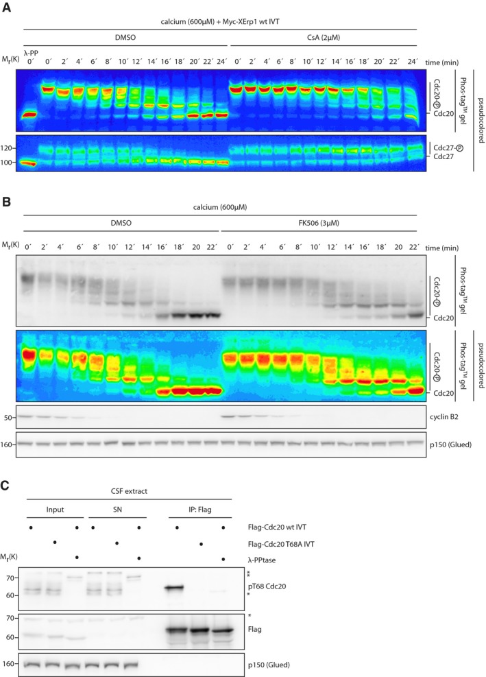
CSF extract was supplemented with Myc‐XErp1 wt IVT and treated with DMSO or CsA. Calcium was added, samples were taken at the indicated time points, resolved by either conventional or Phos‐tag™ SDS–PAGE and immunoblotted for Cdc20, Cdc27, and cyclin B2. A control sample was treated with λ‐phosphatase. A pseudocolored representation of the Cdc20 and the Cdc27 immunoblots is shown in Fig EV4A. The cyclin B2 membrane was stripped and reprobed for α‐tubulin.
CSF extract was supplemented with Flag‐Cdc20 wt IVT coupled to α‐Flag beads. Calcium was added, and Flag‐Cdc20 was isolated by α‐Flag immunoprecipitation at the indicated time points. Input and pellet (IP: Flag) samples were immunoblotted for the Flag‐tag, pThr68 Cdc20 and p150(Glued). The pThr68 Cdc20 membrane was stripped and reprobed for cyclin B2. Asterisk in the cyclin B2 immunoblot indicates the IgG heavy chain. Asterisks in other immunoblots indicate unspecific bands.
CSF extract was supplemented with Flag‐Cdc20 wt IVT. Flag‐Cdc20 was re‐isolated by α‐Flag immunoprecipitation, supplemented with calcium and treated with recombinant hs_CaN and calmodulin as indicated. Samples were taken at the indicated time points and immunoblotted for the Flag‐tag and pThr68 Cdc20. The pThr68 Cdc20 membrane was stripped and reprobed for the catalytic calcineurin subunit CnA. Asterisk indicates the IgG heavy chain.
Pseudocolor representation of the Cdc20 Phos‐tag™ and the Cdc27 immunoblots shown in Fig 3B.
CSF extract was treated with DMSO or FK506. Calcium was added, and samples were taken at the indicated time points. Samples were resolved by either conventional or Phos‐tag™ SDS–PAGE and immunoblotted for Cdc20, cyclin B2, and p150(Glued). A pseudocolored representation of the Cdc20 Phos‐tag™ immunoblot is shown.
CSF extract was supplemented with Flag‐Cdc20 IVT that was either wild‐type or mutated to alanine at Thr68. Flag‐Cdc20 was re‐isolated by α‐Flag immunoprecipitation. Samples and immunoprecipitates were treated with λ‐phosphatase as indicated. Input, supernatant (SN), and pellet (IP: Flag) samples were immunoblotted for the Flag‐tag, pThr68 Cdc20, and p150 (Glued). Asterisks indicate unspecific bands.
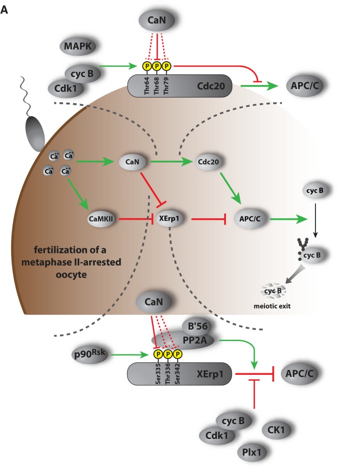
Working model depicting the function of CaMKII and CaN in APC/C activation at meiotic exit in Xenopus laevis oocytes. Green and red connections indicate activating and inhibitory, respectively, relationships.
References
-
- Gross SD, Schwab MS, Taieb FE, Lewellyn AL, Qian YW, Maller JL (2000) The critical role of the MAP kinase pathway in meiosis II in Xenopus oocytes is mediated by p90(Rsk). Curr Biol 10: 430–438 - PubMed
-
- Masui Y, Markert CL (1971) Cytoplasmic control of nuclear behavior during meiotic maturation of frog oocytes. J Exp Zool 177: 129–145 - PubMed
-
- Inoue D, Ohe M, Kanemori Y, Nobui T, Sagata N (2007) A direct link of the Mos‐MAPK pathway to Erp1/Emi2 in meiotic arrest of Xenopus laevis eggs. Nature 446: 1100–1104 - PubMed
Publication types
MeSH terms
Substances
Grants and funding
LinkOut - more resources
Full Text Sources
Miscellaneous
