

Cell-free DNA release under psychosocial and physical stress conditions
- PMID: 30374018
- PMCID: PMC6206142
- DOI: 10.1038/s41398-018-0264-x
Cell-free DNA release under psychosocial and physical stress conditions
Abstract
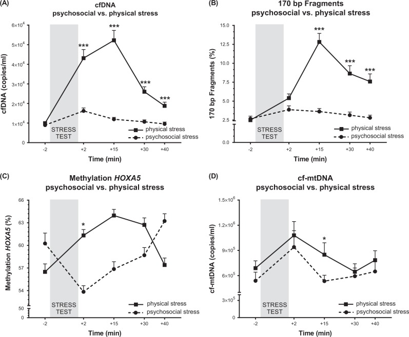
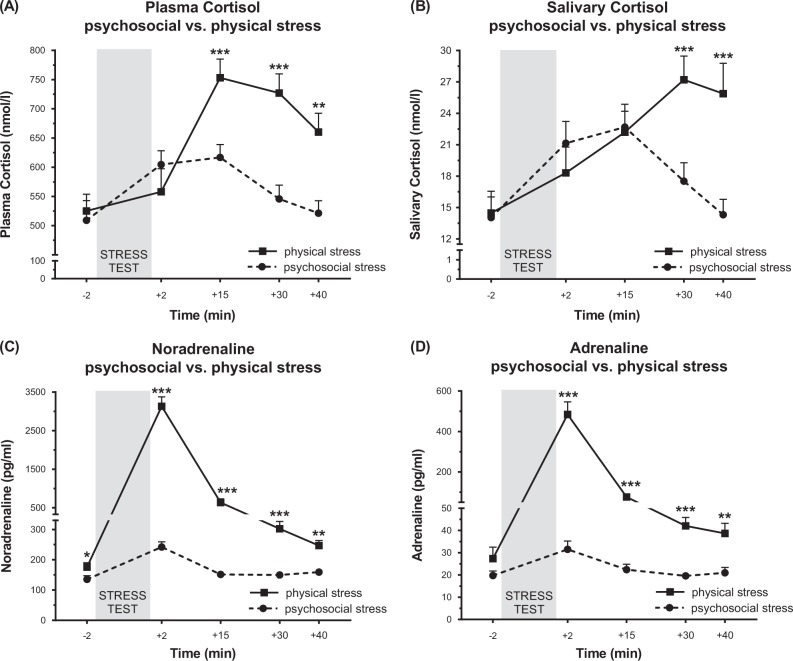
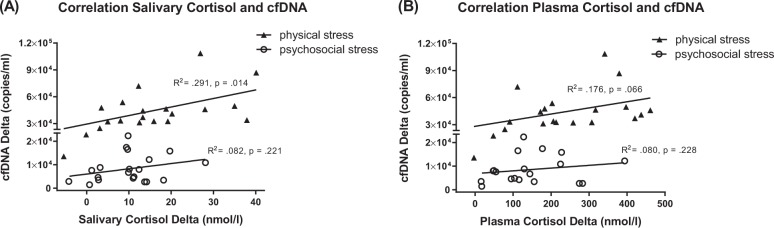
The understanding of mechanisms linking psychological stress to disease risk depend on reliable stress biomarkers. Circulating cell-free DNA (cfDNA) has emerged as a potential biomarker of cellular stress, aging, inflammatory processes, and cell death. Recent studies indicated that psychosocial stress and physical exercise might also influence its release. We compared the effects of acute psychosocial and physical exercise stress on cfDNA release by exposing 20 young, healthy men to both an acute psychosocial laboratory stressor and an acute physical exercise stressor. Venous blood and saliva samples were collected before and after stress exposure. Cell-free DNA was extracted from plasma and quantified by qPCR. Furthermore, cfDNA fragment length was analyzed and cfDNA methylation patterns were assayed across time. In addition, release of stress hormones and subjective stress responses were measured. Results showed a twofold increase of cfDNA after TSST and fivefold increase after exhaustive treadmill exercise, with an overabundance of shorter cfDNA fragments after physical exhaustion. Interestingly, cell-free mitochondrial DNA showed similar increase after both stress paradigms. Furthermore, cfDNA methylation signatures-used here as a marker for diverse cellular origin-were significantly different post stress tests. While DNA methylation decreased immediately after psychosocial stress, it increased after physical stress, suggesting different cellular sources of active DNA release. In summary, our results suggest stimulus and cell-specific regulation of cfDNA release. Whereas the functional role of stress-associated cfDNA release remains elusive, it might serve as a valuable biomarker in molecular stress research as a part of the psychophysiological stress response.
Conflict of interest statement
The authors declare that they have no conflict of interest.
Figures
Similar articles
-
Cell-free DNA release following psychosocial and physical stress in women and men.Transl Psychiatry. 2025 Jan 25;15(1):26. doi: 10.1038/s41398-025-03242-5. Transl Psychiatry. 2025. PMID: 39863589 Free PMC article.
-
Acute high-intensity interval exercise induces comparable levels of circulating cell-free DNA and Interleukin-6 in obese and normal-weight individuals.Life Sci. 2018 Jun 1;202:161-166. doi: 10.1016/j.lfs.2018.04.007. Epub 2018 Apr 10. Life Sci. 2018. PMID: 29653118
-
Exploring the Potential of Cell-Free-DNA Measurements After an Exhaustive Cycle-Ergometer Test as a Marker for Performance-Related Parameters.Int J Sports Physiol Perform. 2017 May;12(5):597-604. doi: 10.1123/ijspp.2016-0157. Epub 2016 Sep 6. Int J Sports Physiol Perform. 2017. PMID: 27617485
-
Circulating cell-free DNA: an up-coming molecular marker in exercise physiology.Sports Med. 2012 Jul 1;42(7):565-86. doi: 10.2165/11631380-000000000-00000. Sports Med. 2012. PMID: 22694348 Review.
-
The diverse origins of circulating cell-free DNA in the human body: a critical re-evaluation of the literature.Biol Rev Camb Philos Soc. 2018 Aug;93(3):1649-1683. doi: 10.1111/brv.12413. Epub 2018 Apr 14. Biol Rev Camb Philos Soc. 2018. PMID: 29654714 Review.
Cited by
-
Liquid biopsies: donor-derived cell-free DNA for the detection of kidney allograft injury.Nat Rev Nephrol. 2021 Sep;17(9):591-603. doi: 10.1038/s41581-021-00428-0. Epub 2021 May 24. Nat Rev Nephrol. 2021. PMID: 34031575 Review.
-
Plasma circulating cell-free mitochondrial DNA in depressive disorders.PLoS One. 2021 Nov 4;16(11):e0259591. doi: 10.1371/journal.pone.0259591. eCollection 2021. PLoS One. 2021. PMID: 34735532 Free PMC article.
-
Healthy mitochondrial DNA in balanced mitochondrial dynamics: A potential marker for neuro‑aging prediction (Review).Biomed Rep. 2023 Aug 7;19(3):64. doi: 10.3892/br.2023.1646. eCollection 2023 Sep. Biomed Rep. 2023. PMID: 37614983 Free PMC article. Review.
-
Deep rest: An integrative model of how contemplative practices combat stress and enhance the body's restorative capacity.Psychol Rev. 2024 Jan;131(1):247-270. doi: 10.1037/rev0000453. Epub 2023 Dec 25. Psychol Rev. 2024. PMID: 38147050 Free PMC article.
-
Circulating Mitochondrial DNA Is Associated With High Levels of Fatigue in Two Independent Sarcoidosis Cohorts.Chest. 2024 May;165(5):1174-1185. doi: 10.1016/j.chest.2023.11.020. Epub 2023 Nov 15. Chest. 2024. PMID: 37977267 Free PMC article.
References
Publication types
MeSH terms
Substances
LinkOut - more resources
Full Text Sources
Other Literature Sources
Medical
