Lipopolysaccharide shock reveals the immune function of indoleamine 2,3-dioxygenase 2 through the regulation of IL-6/stat3 signalling
- PMID: 30374077
- PMCID: PMC6206095
- DOI: 10.1038/s41598-018-34166-4
Lipopolysaccharide shock reveals the immune function of indoleamine 2,3-dioxygenase 2 through the regulation of IL-6/stat3 signalling
Abstract
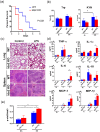
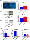
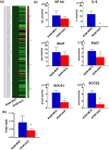
Indoleamine 2,3-dioxygenase 2 (Ido2) is a recently identified catalytic enzyme in the tryptophan-kynurenine pathway that is expressed primarily in monocytes and dendritic cells. To elucidate the biological role of Ido2 in immune function, we introduced lipopolysaccharide (LPS) endotoxin shock to Ido2 knockout (Ido2 KO) mice, which led to higher mortality than that in the wild type (WT) mice. LPS-treated Ido2 KO mice had increased production of inflammatory cytokines (including interleukin-6; IL-6) in serum and signal transducer and activator of transcription 3 (stat3) phosphorylation in the spleen. Moreover, the peritoneal macrophages of LPS-treated Ido2 KO mice produced more cytokines than did the WT mice. By contrast, the overexpression of Ido2 in the murine macrophage cell line (RAW) suppressed cytokine production and decreased stat3 expression. Finally, RAW cells overexpressing Ido2 did not alter nuclear factor κB (NF-κB) or stat1 expression, but IL-6 and stat3 expression decreased relative to the control cell line. These results reveal that Ido2 modulates IL-6/stat3 signalling and is induced by LPS, providing novel options for the treatment of immune disorders.
Conflict of interest statement
The authors declare no competing interests.
Figures


Similar articles
-
Indoleamine 2,3-Dioxygenase 2 Deficiency Exacerbates Imiquimod-Induced Psoriasis-Like Skin Inflammation.Int J Mol Sci. 2020 Aug 1;21(15):5515. doi: 10.3390/ijms21155515. Int J Mol Sci. 2020. PMID: 32752186 Free PMC article.
-
Kynurenine produced by indoleamine 2,3-dioxygenase 2 exacerbates acute liver injury by carbon tetrachloride in mice.Toxicology. 2020 May 30;438:152458. doi: 10.1016/j.tox.2020.152458. Epub 2020 Apr 11. Toxicology. 2020. PMID: 32289347
-
Blockade of indoleamine 2,3-dioxygenase protects mice against lipopolysaccharide-induced endotoxin shock.J Immunol. 2009 Mar 1;182(5):3146-54. doi: 10.4049/jimmunol.0803104. J Immunol. 2009. PMID: 19234212
-
Indoleamine 2,3-dioxygenase-2; a new enzyme in the kynurenine pathway.Int J Biochem Cell Biol. 2009 Mar;41(3):467-71. doi: 10.1016/j.biocel.2008.01.005. Epub 2008 Jan 11. Int J Biochem Cell Biol. 2009. PMID: 18282734 Review.
-
The Two Sides of Indoleamine 2,3-Dioxygenase 2 (IDO2).Cells. 2024 Nov 16;13(22):1894. doi: 10.3390/cells13221894. Cells. 2024. PMID: 39594642 Free PMC article. Review.
Cited by
-
The role of indoleamine 2,3 dioxygenase 1 in the osteoarthritis.Am J Transl Res. 2020 Jun 15;12(6):2322-2343. eCollection 2020. Am J Transl Res. 2020. PMID: 32655775 Free PMC article. Review.
-
Quercetin Administration Suppresses the Cytokine Storm in Myeloid and Plasmacytoid Dendritic Cells.Int J Mol Sci. 2021 Aug 3;22(15):8349. doi: 10.3390/ijms22158349. Int J Mol Sci. 2021. PMID: 34361114 Free PMC article.
-
Current Challenges for IDO2 as Target in Cancer Immunotherapy.Front Immunol. 2021 Apr 21;12:679953. doi: 10.3389/fimmu.2021.679953. eCollection 2021. Front Immunol. 2021. PMID: 33968089 Free PMC article. Review.
-
Impact of IDO1 and IDO2 on the B Cell Immune Response.Front Immunol. 2022 Apr 13;13:886225. doi: 10.3389/fimmu.2022.886225. eCollection 2022. Front Immunol. 2022. PMID: 35493480 Free PMC article. Review.
-
Metabolic reprograming of LPS-stimulated human lung macrophages involves tryptophan metabolism and the aspartate-arginosuccinate shunt.PLoS One. 2020 Apr 8;15(4):e0230813. doi: 10.1371/journal.pone.0230813. eCollection 2020. PLoS One. 2020. PMID: 32267860 Free PMC article.
References
-
- Takikawa O, Yoshida R, KIdo R, Hayaishi O. Tryptophan degradation in mice initiated by indoleamine 2,3-dioxygenase. Journal of Biological Chemistry. 1986;261:3648–3653. - PubMed
-
- Fallarino Francesca, Grohmann Ursula, Vacca Carmine, Orabona Ciriana, Spreca Antonio, Fioretti Maria C., Puccetti Paolo. Advances in Experimental Medicine and Biology. Boston, MA: Springer US; 2003. T Cell Apoptosis by Kynurenines; pp. 183–190. - PubMed
Publication types
MeSH terms
Substances
Grants and funding
LinkOut - more resources
Full Text Sources
Molecular Biology Databases
Research Materials
Miscellaneous

