Review
. 2018 Nov;25(11):1009-1018.
doi: 10.1038/s41594-018-0148-z. Epub 2018 Oct 29.

Organellar TRP channels

Affiliations
Review

Organellar TRP channels

Xiaoli Zhang et al. Nat Struct Mol Biol. 2018 Nov.

Abstract

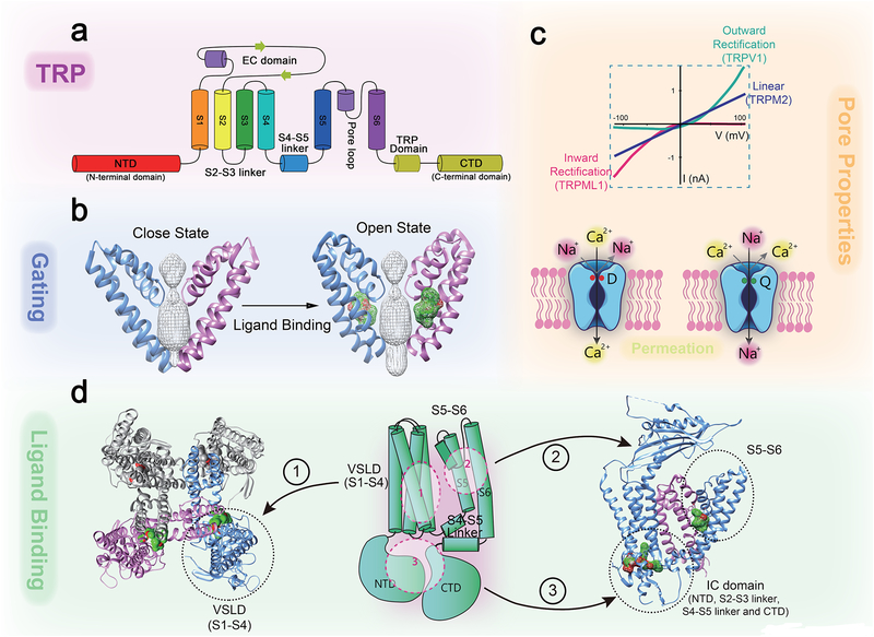
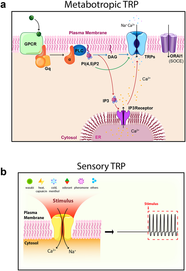
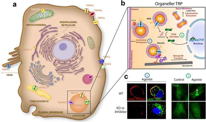
Mammalian transient receptor potential (TRP) channels mediate Ca2+ flux and voltage changes across membranes in response to environmental and cellular signals. At the plasma membrane, sensory TRPs act as neuronal detectors of physical and chemical environmental signals, and receptor-operated (metabotropic) TRPs decode extracellular neuroendocrine cues to control body homeostasis. In intracellular membranes, such as those in lysosomes, organellar TRPs respond to compartment-derived signals to control membrane trafficking, signal transduction, and organelle function. Complementing mouse and human genetics and high-resolution structural approaches, physiological studies employing natural agonists and synthetic inhibitors have become critical in resolving the in vivo functions of metabotropic, sensory, and organellar TRPs.


Figures

Fig. 1.
Fig. 1.. Architecture and functional elements of a TRP channel.
Structural biology analyses reveal critical domains and amino acid residues for Ca2+ permeation and selectivity, and allow visualization of elements such as selectivity filter, activation gates, agonist binding sites, and explain gating-coupling machinery. a. TRPs are 6 transmembrane (TM, S1–S6) cation channels with N- and C- terminal domains facing the cytosol. b, Ligand binding to the S5–S6 domain leads to opening the lower S6 gate. c. Pore properties of TRP channels. Upper panel: representative TRP current-voltage (I-V) traces. Lower panels: the pore-loop between S5 and S6 forms the selectivity filter and upper gate of the channel. Negatively charged residues (e.g. Asp541 in TRPV6; red, the left panel) form the high-affinity Ca2+-binding sites required for Ca2+ permeation and selectivity. In TRPs with very low PCa, specific neutral and polar amino acid residues (e.g., Gln977 in TRPM4; green, the righ panel) form binding sites that favor monovalent cations over Ca2+. d. Ligand binding sites are localized in S1–4 VSLD (site 1), S5–S6 region (site 2; see agonist-bound cryo-EM structures of TRP in the zoomed-in image), or intracellular (IC) domains (site 3; see agonist-bound cryo-EM structures of TRP in the zoomed-in image). The S4–S5 linker and TRP domain act as the binding-gating coupling machinery that interacts to pull the S6 gate open upon ligand-binding.
Fig 2.
Fig 2.. Metabotropic and sensory TRPs.
a. Metabotropic TRPs couple extracellular cues to biology. Excellular neuroendocrine signals, e. g., neurotransmitters, act on G protein-coupled receptors (GPCRs). Metabotropic TRPs are signal transducers in cells that also express phospholipase (PLC)-dependent GPCRs. Activation of Gq-coupled receptors stimulates PLC activity, which hydrolyze PI(4,5)P2 into DAG and IP3. IP3 then induces Ca2+ release from the ER through IP3 receptors. PLC-dependent signal transduction mechanisms activate metabotropic TRPs, as well as store-operated Ca2+ entry channels, whose pore-forming subunits are Orai proteins. Details see Table 1. b. Sensory TRPs couple environmental cues to biology. Sensory TRPs are activated by environmental signals such as light, temperature change, osmo-mechanical force, and plant-derived compounds, pain-/itch-inducing chemicals, tastants, and pheromones. These physical and chemical signals activate sensory TRP-mediated Na+ entry, increasing the membrane excitability of various sensory cells, including DRG neurons, taste receptor cells, hair cells, and retinal ganglion cells through sensory TRPs. Details see Table 1.
Fig. 3.
Fig. 3.. Organellar TRP channels.
a. Though most TRPs are located at the plasma membrane, TRPMLs are localized in intracellular endosomes and lysosomes, and TRPPs are localized in primary cilia. In addition, TRPV1 is localized in the ER and possibly mitochondria, and TRPM7 is localized in M7-like vesicles. In specialized cell types, TRPA1 and TRPM2 are functionally expressed in lysosomes. In parietal cells, TRPML1 is an organellar TRP that functions in both lysosomes and tubulovesicles (TVs). b. Endogenous (e.g. ROS and PI(3,5)P2) or synthetic agonists induce TRPML1-mediated lysosomal Ca2+ release to trigger lysosomal exocytosis (example 1), retrograde transport, and TFEB nuclear translocation (example 2). In the parietal cells, histamine-induced cAMP/protein kinase A signaling activates TV-localized TRPML1, increasing TV trafficking and exocytosis. c. In example 1, activation of TRPML1 triggers lysosomal exocytosis, detected by the surface expression of Lamp1 proteins in WT, but not in TRPML1 KO cells. Images are modified with permission from ref.; In example 2, agonist activation of TRPML1 leads to nuclear translocation of TFEB (green), a transcription factor for lysosome biogenesis and autophagy in WT, but not TRPML1 KO cells. Images are modified with permission from ref. 102.
Box Fig. 1.
Box Fig. 1.. An integrated approach to study TRPs.
An integrated approach to establish functions of TRPs with respect to channel physiology, cell and tissue physiology, and organismal biology, is illustrated below, in anticlockwise direction. Top right, agonist-evoked inward currents (measured at negative voltages) are abolished in TRP KO cells or by synthetic inhibitors, suggestive of the specificity of the response. Bottom right, structural biology analyses reveal critical domains and amino acid residues as agonist binding sites. Middle right, corresponding mutations in the binding sites abolished agonist-induced Ca2+ imaging responses. Bottom left, cell biological functions of TRPs (e.g. agonist activation of TRPML1 leads to nuclear translocation of transcription factor TFEB in WT, but not KO cells). Top left, animal biology and physiology of TRPs. For example, agonist (e.g., capsaicin for TRPV1) application increases firing frequency in WT, but not TRP KO, DRG neurons.

References

    1. Minke B, Wu C & Pak WL Induction of photoreceptor voltage noise in the dark in Drosophila mutant. Nature 258, 84–87 (1975). - PubMed
    1. Montell C & Rubin GM Molecular characterization of the Drosophila trp locus: a putative integral membrane protein required for phototransduction. Neuron 2, 1313–1323 (1989). - PubMed
    1. Hardie RC & Minke B The trp gene is essential for a light-activated Ca2+ channel in Drosophila photoreceptors. Neuron 8, 643–651 (1992). - PubMed
    1. Wes PD et al. TRPC1, a human homolog of a Drosophila store-operated channel. Proc Natl Acad Sci U S A 92, 9652–9656 (1995). - PMC - PubMed
    1. Zhu X, Chu PB, Peyton M & Birnbaumer L Molecular cloning of a widely expressed human homologue for the Drosophila trp gene. FEBS Lett 373, 193–198 (1995). - PubMed

Publication types

Substances