. 2018 Dec;14(12):1127-1132.
doi: 10.1038/s41589-018-0153-x. Epub 2018 Oct 29.

The multicatalytic compartment of propionyl-CoA synthase sequesters a toxic metabolite

Affiliations

The multicatalytic compartment of propionyl-CoA synthase sequesters a toxic metabolite

Iria Bernhardsgrütter et al. Nat Chem Biol. 2018 Dec.

Abstract

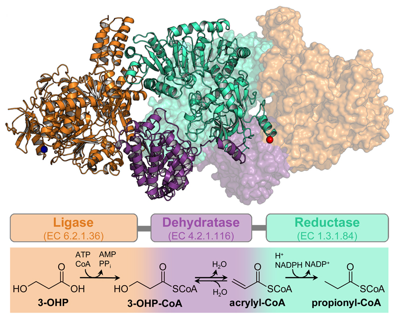
Cells must cope with toxic or reactive intermediates formed during metabolism. One coping strategy is to sequester reactions that produce such intermediates within specialized compartments or tunnels connecting different active sites. Here, we show that propionyl-CoA synthase (PCS), an ∼ 400-kDa homodimer, three-domain fusion protein and the key enzyme of the 3-hydroxypropionate bi-cycle for CO2 fixation, sequesters its reactive intermediate acrylyl-CoA. Structural analysis showed that PCS forms a multicatalytic reaction chamber. Kinetic analysis suggested that access to the reaction chamber and catalysis are synchronized by interdomain communication. The reaction chamber of PCS features three active sites and has a volume of only 33 nm3. As one of the smallest multireaction chambers described in biology, PCS may inspire the engineering of a new class of dynamically regulated nanoreactors.


Conflict of interest statement

Competing interests The authors declare no competing financial interests.

Figures

Figure 1
Figure 1. Trifunctional PCS: Structure and reaction sequence.
Dimeric structure of PCS from Erythrobacter sp. NAP1 (PDB 6EQO). One protomer is depicted in cartoon and one in surface representation. The multi-domain organization is highlighted by different colors: orange, ligase domain; purple, dehydratase domain; cyan, reductase domain; blue sphere, N-terminus; red sphere, C-terminus. Schematic arrangement of the three domains and their individual reactions are shown using the same color code.
Figure 2
Figure 2. PCS sequesters the reactive intermediate acrylyl-CoA.
a, Time course of the overall reaction with 0.1 µM PCS, 800 µM CoA, 500 µM 3-hydroxypropionate, 800 µM ATP and 300 µM NADPH. Production of the 3-hydroxypropionyl-CoA intermediate (orange) and the final product propionyl-CoA (cyan) was observed. In contrast no free acrylyl-CoA was detectable. b, Time course of the reaction containing 10 µM PCS, 5 mM CoA, 5 mM 3-hydroxypropionate, 5 mM ATP and 5 mM NADPH. At these high enzyme concentrations acrylyl-CoA (purple) was detected at 0.18 µM during steady-state corresponding to 1.8% occupancy of reductase active sites. 3-Hydroxypropionyl-CoA und propionyl-CoA accumulate over time. c, as in a, but without NADPH. Again, formation of 3-hydroxypropionyl-CoA was observed, but not of free acrylyl-CoA. d, Isotopic labeling competition experiment containing unlabeled 3-hydroxypropionate and either 13C-labeled 3-hydroxypropionyl-CoA (experiment 1) or acrylyl-CoA (experiment 2). The reaction was started by the addition of PCS. Products were analyzed by LC-MS (see Supplementary Table 4 for detailed assay conditions). e, Results of the isotopic labeling competition experiment. Only 0.8 ± 0.4 % of propionyl-CoA was produced from exogenous 13C-labeled 3-hydroxypropionyl-CoA during steady state (experiment 1). Approximately every fifth propionyl-CoA (21 ± 15%) was formed from exogenous 13C-labeled acrylyl-CoA during steady-state (experiment 2). ac, data of a representative single experiment. e, data mean ± s.d. (n=3).
Figure 3
Figure 3. Multi-catalytic reaction chamber of PCS.
Volume filling representation of the reaction chambers enclosed by PCS. The central catalytic reaction chamber of each protomer is formed through the contribution of all three domains. Orange, contribution of the ligase domain; purple, contribution of the dehydratase domain; cyan, contribution of the reductase domain. The close up shows a cross section through the reaction chamber. Electrostatic charge distribution is shown as a gradient from negatively charged (red) to positively charged (blue). The three active sites are well connected within the reaction chamber. Large positively charged patches may help in retaining the CoA-ester intermediates inside during catalysis or even guide them between active sites. Negative charges around the small openings may also prevent leakage of the negatively charged CoA-derivatives. The PCS structure co-crystallized with CoA (no density), an ATP analog and NADP+ is depicted in cartoon showing the ligase domain in orange, the dehydratase domain in purple and the reductase domain in cyan. CoA binding sites have been modelled based on the superposition of the structures of lone-standing CoA ligase (PDB 2P2F), dehydratase (PDB 5JBX) and reductase (PDB 4A0S) onto PCS (compare Supplementary Figs. 6, 7 and 8). Distances between the active sites have been determined by measuring the distance between sulfur atoms of modelled CoA moieties: ligase–dehydratase, 42.5 Å; dehydratase–reductase, 33.7 Å; ligase–reductase, 63.5 Å.
Figure 4
Figure 4. Proposed catalytic cycle of PCS.
In the open conformation, 3-hydroxypropionate (3OHP) and ATP are converted to 3-hydroxypropionyl-AMP (3OHP-AMP) through the ligase domain (orange). The binding of CoA induces closing of the enzyme and the formation of 3-hydroxypropionyl-CoA (3OHP-CoA). 3OHP-CoA is released into the reaction chamber, where it is converted by the dehydratase domain (purple) to acrylyl-CoA. Acrylyl-CoA then enters the active site of the reductase domain (cyan). Following the reduction of acrylyl-CoA to propionyl-CoA, the reaction chamber reopens to release propionyl-CoA, which leaves PCS ready for the next catalytic cycle.

Similar articles

Cited by

References

    1. Wheeldon I, et al. Substrate channelling as an approach to cascade reactions. Nat Chem. 2016;8:299–309. - PubMed
    1. Linster CL, Van Schaftingen E, Hanson AD. Metabolite damage and its repair or pre-emption. Nat Chem Biol. 2013;9:72–80. - PubMed
    1. Wheeldon I, et al. Substrate channelling as an approach to cascade reactions. Nature chemistry. 2016;8:299. - PubMed
    1. Alber BE, Fuchs G. Propionyl-coenzyme A synthase from Chloroflexus aurantiacus, a key enzyme of the 3-hydroxypropionate cycle for autotrophic CO2 fixation. J Biol Chem. 2002;277:12137–12143. - PubMed
    1. Zarzycki J, Brecht V, Müller M, Fuchs G. Identifying the missing steps of the autotrophic 3-hydroxypropionate CO2 fixation cycle in Chloroflexus aurantiacus. Proc Natl Acad Sci USA. 2009;106:21317–21322. - PMC - PubMed

Publication types

MeSH terms