. 2018 Oct 24:7:79.
doi: 10.1038/s41377-018-0080-3. eCollection 2018.

Optical prediction of single muscle fiber force production using a combined biomechatronics and second harmonic generation imaging approach

Affiliations

Optical prediction of single muscle fiber force production using a combined biomechatronics and second harmonic generation imaging approach

Dominik Schneidereit et al. Light Sci Appl. .

Abstract

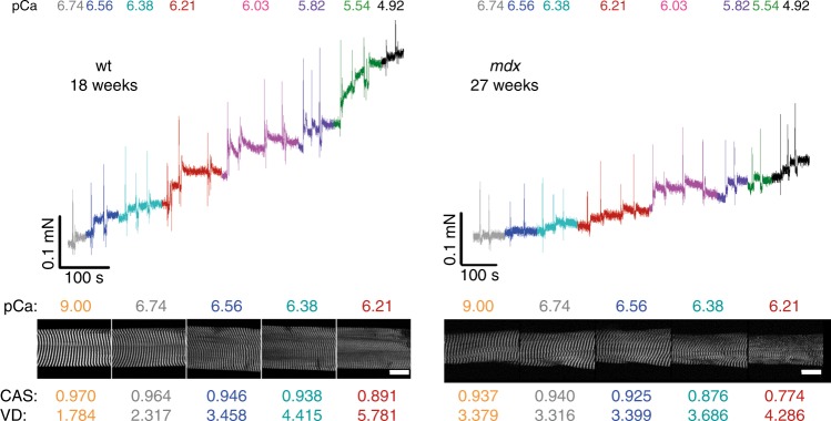
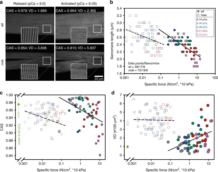
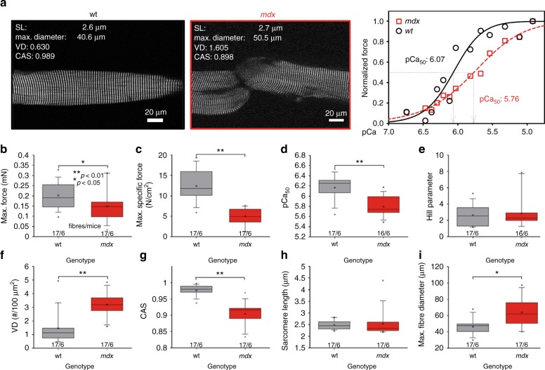
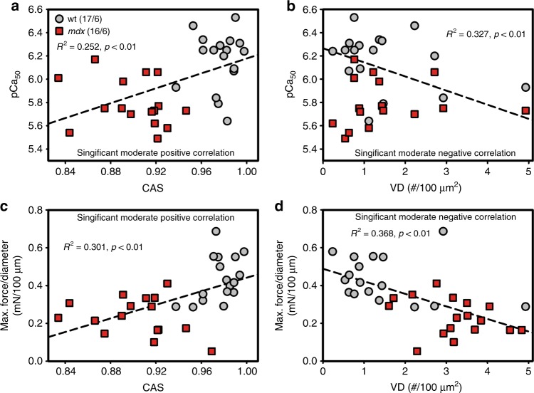
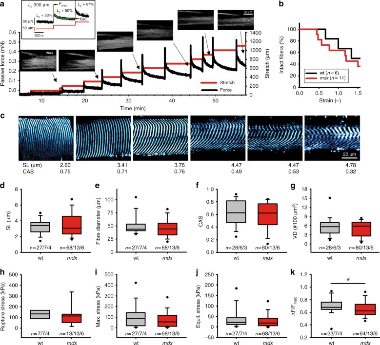
Skeletal muscle is an archetypal organ whose structure is tuned to match function. The magnitude of order in muscle fibers and myofibrils containing motor protein polymers determines the directed force output of the summed force vectors and, therefore, the muscle's power performance on the structural level. Structure and function can change dramatically during disease states involving chronic remodeling. Cellular remodeling of the cytoarchitecture has been pursued using noninvasive and label-free multiphoton second harmonic generation (SHG) microscopy. Hereby, structure parameters can be extracted as a measure of myofibrillar order and thus are suggestive of the force output that a remodeled structure can still achieve. However, to date, the parameters have only been an indirect measure, and a precise calibration of optical SHG assessment for an exerted force has been elusive as no technology in existence correlates these factors. We engineered a novel, automated, high-precision biomechatronics system into a multiphoton microscope allows simultaneous isometric Ca2+-graded force or passive viscoelasticity measurements and SHG recordings. Using this MechaMorph system, we studied force and SHG in single EDL muscle fibers from wt and mdx mice; the latter serves as a model for compromised force and abnormal myofibrillar structure. We present Ca2+-graded isometric force, pCa-force curves, passive viscoelastic parameters and 3D structure in the same fiber for the first time. Furthermore, we provide a direct calibration of isometric force to morphology, which allows noninvasive prediction of the force output of single fibers from only multiphoton images, suggesting a potential application in the diagnosis of myopathies.


Figures

Fig. 1
Fig. 1. A novel biomechatronics system (MechaMorph) for the simultaneous assessment of isometric, Ca2+-activated force and SHG multiphoton imaging in single muscle fibers.
a 3D CAD sketch of the miniaturized biomechatronics device containing an interchangeable muscle fiber chamber to fit onto a stage of a multiphoton microscope. A force transducer and voice coil actuator pin are connected to a horizontal trough, each fabricated from cannula needles to take up a single muscle fiber (d). b photograph of the engineered device which is inserted in between two objective lenses to record forward scattered SHG, while the custom-made biomechatronics software runs on a parallel computer (c, d)
Fig. 2
Fig. 2. Ca2+-activated force and SHG multiphoton imaging simultaneously performed in single wt and mdx EDL muscle fibers.
Representative example recordings of force (top) and myosin SHG signals (bottom) from a single EDL fiber from a wt mouse (left) and an mdx mouse (right) during successive solution exchange for increasing Ca2+ concentrations (decreasing pCa). Brief positive force spikes represent the time points of manual solution exchange performed several times per pCa step. Scale bar: 20 µm. CAS: cosine angle sum (a.u.). VD: vernier density (#/100 µm2)
Fig. 3
Fig. 3. Distortion of sarcomeric and myofibrillar structures in single EDL muscle fibers from both wt and mdx mice during active Ca2+-induced isometric force generation.
a representative example SHG images of a single EDL fiber from a wt mouse (17 weeks) and an mdx mouse (27 weeks) in the relaxed pCa 9 and pCa 6.03 activated states. During mechanical activation, although isometric, the sarcomere lengths visibly shorten and fiber diameters increase. b analysis of the reduction in single fiber sarcomere length during Ca2+-dependent force generation (logarithmic plot of specific force) in wt mice (filled circles) and mdx mice (open squares); pCa values color-coded. c, d, changes in myofibrillar/sarcomeric ultrastructural parameters of the cosine angle sum, CAS (c), and vernier density, VD (d), indicate a Ca2+-graded increase in the myofibrillar disorder (CAS↓, VD↑) that is more pronounced in the ordered wt EDL fibers, while single fibers from mdx mice are already highly disordered under relaxed conditions
Fig. 4
Fig. 4. Myofibrillar ultrastructural disorganization in relaxed mdx EDL fibers is a predictor of reduced contractile performance and reduced Ca2+ sensitivity of the contractile apparatus.
a SHG images of single EDL fibers from a wt mouse (14 weeks) and an mdx mouse (79 weeks) in the relaxed state (pCa 9) with indicated sarcomere lengths (SL), maximum diameters, VD and CAS. Steady-state force at a given pCa normalized to the maximum force with indicated pCa50 and Hill parameters for the shown fibers. Single mdx EDL fibers showed vast biomechanical deficits in active force production: significantly reduced max. absolute (b) and specific force (c) at a pCa of 4.92–5.67 and significantly reduced pCa50 values indicative of myofibrillar Ca2+ desensitization (d) with otherwise similar Hill parameters (e). This correlates well with myofibrillar structural deficits, as shown by the markedly increased VD (f) and decreased CAS (g) in mdx mice over wt mice. At similar sarcomere lengths (h), fiber diameters were larger in mdx EDL fibers compared than in wt fibers (i). Box plots with box (25 to 75 percentiles), median (line), whiskers (5–95 percentiles), minimum and maximum (x), mean (rectangle) and significances from one-way ANOVA with post hoc Bonferroni test (equal variance) or post hoc Tukey test (no equal variance) indicated as p < 0.05 (*) and p < 0.01 (**)
Fig. 5
Fig. 5. Pearson correlations show significant correlations between SHG-derived structural data and active isometric force parameters in single EDL fibers.
Both structural parameters, the CAS and VD, obtained in relaxed (pCa 9) wt (gray) and mdx (red) single fibers show significant correlations with pCa50 and the maximum force per diameter in the activated state. In particular, the more ordered the myofibrillar arrangement (CAS↑, VD↓), the higher the probability for a higher Ca2+ sensitivity (larger pCa50) (a, b), and thus, a higher predicted force production upon Ca2+ activation (c, d). Thus, the CAS positively correlates with both pCa50 and the max. force (a, c), while VD negatively correlates with both (b, d)
Fig. 6
Fig. 6. Simultaneous assessment of passive single fiber viscoelasticity biomechanics and SHG myofibrillar ultrastructure in wt and mdx EDL muscles.
a sequence of SHG images (left) taken during a protocol stretching a single EDL fiber from an mdx mouse in 50 µm steps and recording restoration force (right). Images were taken at the time points indicated. The inset shows force recording of another mdx fiber: instantaneous maximum restoration force Fmax at each stretch was followed by a double-exponential viscous relaxation to a new steady-state elastic force level Feq. b mdx single EDL fibers already broke at lower strains compared to wt fibers, compatible with larger stiffness, respective lower viscous relaxation. c sequence of SHG images taken from a wt single EDL fiber stretched to the indicated sarcomere lengths (SL) and analyzing CAS values. Note that with the increase in stretch, a marked decline in CAS can be detected as visualized by A-band bending across the fiber cross-section. The SL value ranges were similar in wt and mdx fibers (d), as were the fiber diameters (e), overall CAS values (f) and VDs (g). Biomechanical passive parameters of the maximum rupture stress (h), maximum stress values from all stretches (i) and equilibrium strains (j) were similar among the wt and mdx fibers while the viscous stress relief ΔF/Fmax values (k) were significantly smaller in the mdx fibers than the wt fibers, indicating a lower viscosity in the dystrophic genotype. #P < 0.025. n = (a/b/c) depicts the data from a images on b fibers from c animals
Fig. 7
Fig. 7. Pearson correlations show significant correlations between the SHG-derived structural data and the passive elastic parameters, as well as a higher degree of myofibrillar disorder with the sarcomere length in stretched single EDL fibers.
Both structural parameters, the CAS (a) and the stretch-corrected VD (b), obtained in relaxed (pCa 9) wt (gray) and mdx (red) single fibers show significant correlations with the sarcomere lengths. In particular, the higher the stretch was (SL↑), the less ordered the myofibrillar arrangement became (CAS↓, VD↑). Similarly, the myofibrillar disarray, i.e., the low CAS (c) and large VD values (d), significantly correlated with increased maximum stress values that occurred during stretching

Similar articles

Cited by

References

    1. Clark KA, McElhinny AS, Beckerle MC, Gregorio CC. Striated muscle cytoarchitecture: an intricate web of form and function. Annu. Rev. Cell. Dev. Biol. 2002;18:637–706. doi: 10.1146/annurev.cellbio.18.012502.105840. - DOI - PubMed
    1. Goldfarb LG, Dalakas MC. Tragedy in a heartbeat: malfunctioning desmin causes skeletal and cardiac muscle disease. J. Clin. Invest. 2009;119:1806–1813. doi: 10.1172/JCI38027. - DOI - PMC - PubMed
    1. Both M, et al. Second harmonic imaging of intrinsic signals in muscle fibers in situ. J. Biomed. Opt. 2004;9:882–892. doi: 10.1117/1.1783354. - DOI - PubMed
    1. Rode C, Siebert T, Tomalka A, Blickhan R. Myosin filament sliding through the Z-disc relates striated muscle fibre structure to function. Proc. Biol. Sci. 2016;283:20153030. doi: 10.1098/rspb.2015.3030. - DOI - PMC - PubMed
    1. Berchtold MW, Brinkmeier H, Müntener M. Calcium ion in skeletal muscle: its crucial role for muscle function, plasticity, and disease. Physiol. Rev. 2000;80:1215–1265. doi: 10.1152/physrev.2000.80.3.1215. - DOI - PubMed