. 2018 Oct 22:4:51.
doi: 10.1038/s41420-018-0110-1. eCollection 2018.

Lon protease inactivation in Drosophila causes unfolded protein stress and inhibition of mitochondrial translation

Affiliations

Lon protease inactivation in Drosophila causes unfolded protein stress and inhibition of mitochondrial translation

Gautam Pareek et al. Cell Death Discov. .

Erratum in

Abstract

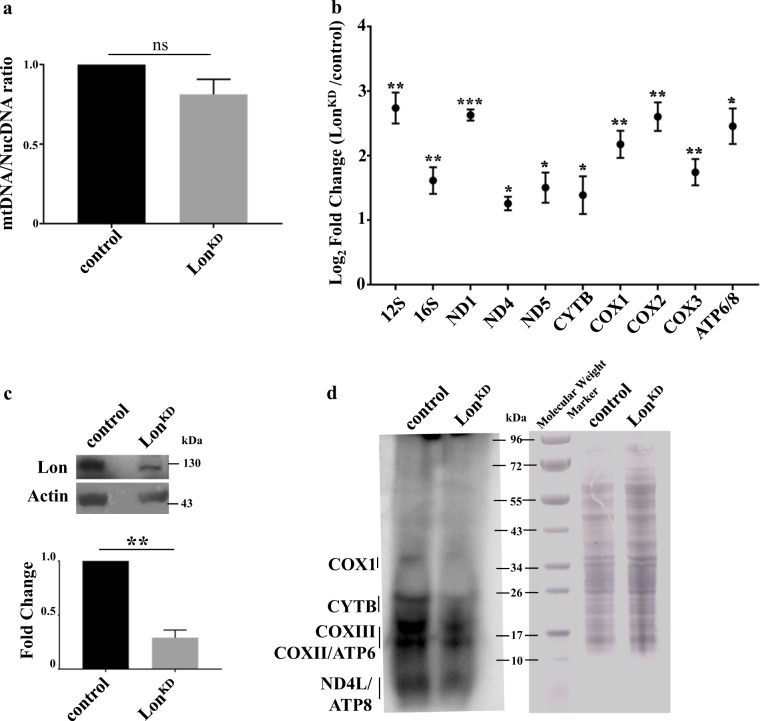
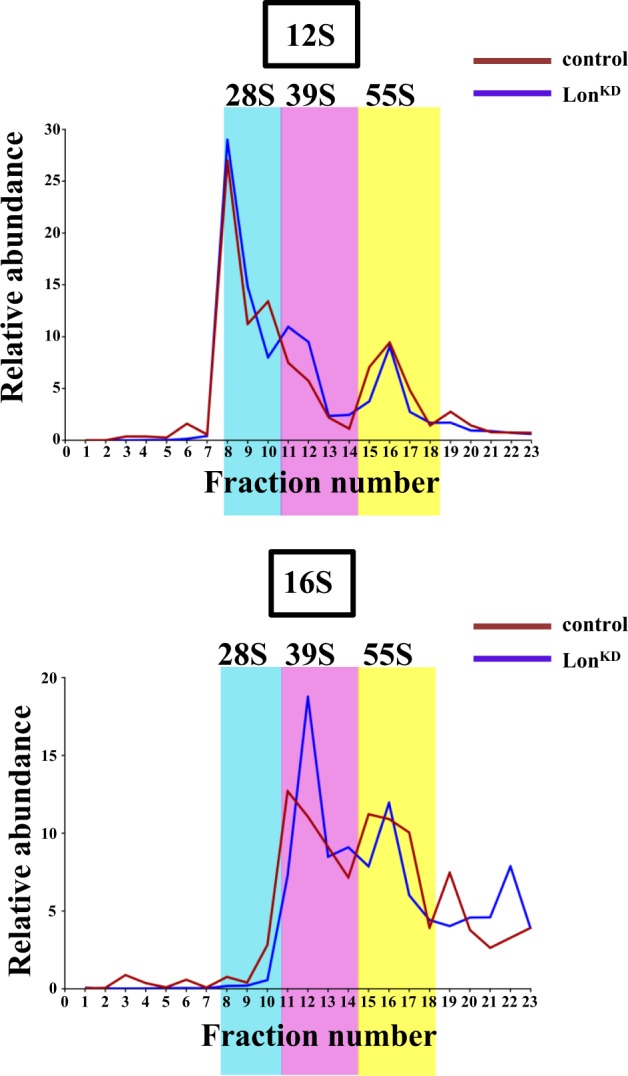
Mitochondrial dysfunction is a frequent participant in common diseases and a principal suspect in aging. To combat mitochondrial dysfunction, eukaryotes have evolved a large repertoire of quality control mechanisms. One such mechanism involves the selective degradation of damaged or misfolded mitochondrial proteins by mitochondrial resident proteases, including proteases of the ATPase Associated with diverse cellular Activities (AAA+) family. The importance of the AAA+ family of mitochondrial proteases is exemplified by the fact that mutations that impair their functions cause a variety of human diseases, yet our knowledge of the cellular responses to their inactivation is limited. To address this matter, we created and characterized flies with complete or partial inactivation of the Drosophila matrix-localized AAA+ protease Lon. We found that a Lon null allele confers early larval lethality and that severely reducing Lon expression using RNAi results in shortened lifespan, locomotor impairment, and respiratory defects specific to respiratory chain complexes that contain mitochondrially encoded subunits. The respiratory chain defects of Lon knockdown (Lon KD ) flies appeared to result from severely reduced translation of mitochondrially encoded genes. This translational defect was not a consequence of reduced mitochondrial transcription, as evidenced by the fact that mitochondrial transcripts were elevated in abundance in Lon KD flies. Rather, the translational defect of Lon KD flies appeared to be derived from sequestration of mitochondrially encoded transcripts in highly dense ribonucleoparticles. The translational defect of Lon KD flies was also accompanied by a substantial increase in unfolded mitochondrial proteins. Together, our findings suggest that the accumulation of unfolded mitochondrial proteins triggers a stress response that culminates in the inhibition of mitochondrial translation. Our work provides a foundation to explore the underlying molecular mechanisms.


Conflict of interest statement

The authors declare that they have no conflict of interest.

Figures

Fig. 1
Fig. 1. Inactivation of Lon results in shortened lifespan and defective locomotion.
a Immunoblot analysis from heads of 1-day-old control and LonKD flies using Lon and actin antibodies. The Lon band intensity is normalized against actin as a loading control. Significance was determined using Student’s t-test (***p< 0.0005 by Student’s t-test). The experiment was repeated at least three times. b Kaplan–Meier survival curves of LonKD flies (n = 167, 50% survival 31 days) and controls (n = 174, 50% survival 73 days) (****p < 0.0001 by Mantel–Cox log-rank test). c One-day-old LonKD flies exhibit a climbing defect. Error bars represent SEM (n= 135 for LonKD, 134 for control, ***p < 0.0005 by Student’s t-test). d One-day-old LonKD flies exhibit a significant decrease in flight index (n= 6 independent groups of 10–15 animals, **p < 0.005 from Student’s t-test). Control = UAS-mCherry-RNAi driven by elav-GAL4 and da-GAL4. LonKD = UAS-Lon-RNAi-1 driven by elav-GAL4 and da-GAL4
Fig. 2
Fig. 2. Lon knockdown flies have altered respiratory chain function and abundance.
a The activity of respiratory chain complexes in control and LonKD flies at 1 day (left panel) and 21 days (right panel) of age (n = 2 independent groups of 500 adult flies, *p < 0.05, **p < 0.005 from Student’s t-test). b BN-PAGE analysis of mitochondrial protein extracts from 21-day-old control and LonKD flies. The red asterisk marks the location of the subcomplex containing F1 subunit of ATP synthase that is only detected in LonKD flies. Immunoblot of citrate synthase (bottom panel) was used as a loading control. c In-gel activity of mitochondrial complexes I and IV isolated from 21-day-old adults. SCs here refer to the supercomplexes. d In-gel activity of mitochondrial complex II isolated from 21-day-old flies. e In-gel activity of mitochondrial complex V isolated from 21-day-old adults. The red asterisk denotes the location of the subcomplex containing Fsubunit of ATP synthase in LonKD flies. Note that the protein molecular weight markers shown in (b) were used as reference to mark the gels in (c–e). For BN-PAGE and in-gel activity assays, the images shown are representative of two independent biological replicates. Control = UAS-mCherry-RNAi driven by elav-GAL4 and da-GAL4. LonKD = UAS-Lon-RNAi-1 driven by elav-GAL4 and da-GAL4
Fig. 3
Fig. 3. Lon knockdown flies exhibit increased mitochondrial transcript abundance and decreased mitochondrial translation.
a Mitochondrial DNA abundance was compared in LonKD and control flies using qPCR to compare the ratio of mtDNA-encoded mt:Cyt-b to that of nuclear-encoded Act79b (n = 3 independent groups of 40–45 fly heads). Statistical significance was determined using Student’s t-test. b qRT-PCR was used to quantify steady-state abundance of the indicated mitochondrial RNAs in 21-day-old adult fly heads. Mitochondrial RNA abundance was normalized to the abundance of the nuclear-encoded Act79b transcript (n = 3 independent groups of 40–45 fly heads). Error bars indicate mean ± SEM. Student’s t-test was applied, *p < 0.05, **p < 0.005, ***p < 0.0005. c Immunoblot analysis of third instar larvae to confirm knockdown of Lon using actin as a loading control. n = 3 independent groups of five third instar larvae. Significance was determined using Student’s t-test, **p < 0.005. d In organello translation was performed using mitochondria isolated from third instar larvae. Mitochondria were labeled by incubating with 35S-methionine for 1 h. Positions of individual mitochondrially encoded proteins are indicated (left panel). Coomassie-stained gel (right panel) was used as a loading control. Control = UAS-mCherry-RNAi driven by elav-GAL4 and da-GAL4. LonKD = UAS-Lon-RNAi-1 driven by elav-GAL4 and da-GAL4
Fig. 4
Fig. 4. Mitochondrial ribosome assembly is only mildly affected in LonKD flies.
Mitochondrial lysates from 21-day-old control and LonKD flies were subjected to sucrose density gradient fractionation to assess the state of assembly of mitochondrial ribosomes. The relative proportions of the small (28S) subunit, large (39S) subunit, and fully assembled (55S) mitochondrial ribosomes were assessed by subjecting the density gradient fractions to qRT-PCR using primer sets specific to 12S rRNA, which marks the small subunit, and to 16S rRNA, which marks the large subunit. Co-localization of the 12S and 16S rRNAs is diagnostic of fully assembled and actively translating ribosomes. Fractions containing the small (28S) subunit, large (39S) subunit, and fully assembled (55S) ribosome are shaded cyan, pink, and yellow, respectively. The relative abundance of a given rRNA in each fraction was calculated as the percentage relative to the total RNA abundance in all fractions after normalizing to a luciferase control RNA that was spiked into each of the fractions prior to RNA isolation. Control = UAS-mCherry-RNAi driven by elav-GAL4 and da-GAL4. LonKD = UAS-Lon-RNAi-1 driven by elav-GAL4 and da-GAL4
Fig. 5
Fig. 5. Mitochondrial RNAs accumulate in large untranslated particles upon Lon knockdown.
qRT-PCR analysis of individual sucrose gradient fractions was used to characterize the distribution of the indicated mtDNA-encoded mRNAs relative to the 28S and 39S subunits, and fully assembled 55S mitochondrial ribosomes. Mitochondrial homogenates for sucrose density fractionation were prepared from 21-day-old adult LonKD and control flies. Relative abundance represents the fraction of the mRNA in any given fraction relative to the total after normalizing to a luciferase control RNA that was added to each of the fractions prior to RNA isolation. Control = UAS-mCherry-RNAi driven by elav-GAL4 and da-GAL4. LonKD = UAS-Lon-RNAi-1 driven by elav-GAL4 and da-GAL4
Fig. 6
Fig. 6. Inactivation of Lon protease results in the accumulation of unfolded mitochondrial proteins.
a Triton-insoluble mitochondrial proteins detected by western blot in heads from 1-day-old control and LonKD flies, using antibodies to complex Vβ, PDHα, aconitase, and NDUFS3. The results were quantified by ratio to actin and normalized to control levels. The experiment was repeated at least three times. *p< 0.05, **p< 0.005, ***p< 0.0005 by Student’s t-test. b Immunoblot analysis of head proteins using antibodies to Hsp60A and Hsc70-5. Protein was extracted from heads of day 1 control and LonKD flies using RIPA buffer. Quantification was performed as in (a). Experiments were repeated at least three times. *p< 0.05, **p< 0.005 by Student’s t-test. Control = UAS-mCherry-RNAi driven by elav-GAL4 and da-GAL4. LonKD = UAS-Lon-RNAi-1 driven by elav-GAL4 and da-GAL4
Fig. 7
Fig. 7. ClpP overexpression rescues the climbing defect of Lon-deficient flies.
a Immunoblot analysis of fly heads to confirm expression of FLAG-tagged ClpP in flies bearing a UAS-ClpP-FLAG-HA transgene and the elav-GAL4 pan-neuronal driver. b Climbing was measured in 1-day-old control flies (n= 94), LonKD-elav flies (n = 91), and LonKD-elav flies co-expressing ClpP protease (n = 83). Control = UAS-mCherry-RNAi driven by elav-GAL4. LonKD-elav; mCherry RNAi=UAS-Lon-RNAi-1 and UAS-mCherry-RNAi driven by elav-GAL4. LonKD-elav; UAS-ClpP = UAS-Lon-RNAi-1 and UAS-ClpP-FLAG-HA driven by elav-GAL4. Error bars represent SEM. ***p< 0.0005, ****p< 0.0001 by Student’s t-test

Similar articles

Cited by

References

    1. Kauppila TES, Kauppila JHK, Larsson NG. Mammalian mitochondria and aging: an update. Cell Metab. 2017;25:57–71. doi: 10.1016/j.cmet.2016.09.017. - DOI - PubMed
    1. Sun N, Youle RJ, Finkel T. The mitochondrial basis of aging. Mol. Cell. 2016;61:654–666. doi: 10.1016/j.molcel.2016.01.028. - DOI - PMC - PubMed
    1. Pickrell AM, Youle RJ. The roles of PINK1, parkin, and mitochondrial fidelity in Parkinson’s disease. Neuron. 2015;85:257–273. doi: 10.1016/j.neuron.2014.12.007. - DOI - PMC - PubMed
    1. Gustafsson CM, Falkenberg M, Larsson NG. Maintenance and expression of mammalian mitochondrial DNA. Annu. Rev. Biochem. 2016;85:133–160. doi: 10.1146/annurev-biochem-060815-014402. - DOI - PubMed
    1. Hallberg BM, Larsson NG. Making proteins in the powerhouse. Cell Metab. 2014;20:226–240. doi: 10.1016/j.cmet.2014.07.001. - DOI - PubMed

LinkOut - more resources