. 2018 Oct 15:8:353.
doi: 10.3389/fcimb.2018.00353. eCollection 2018.

Activity of Thioallyl Compounds From Garlic Against Giardia duodenalis Trophozoites and in Experimental Giardiasis

Affiliations

Activity of Thioallyl Compounds From Garlic Against Giardia duodenalis Trophozoites and in Experimental Giardiasis

Raúl Argüello-García et al. Front Cell Infect Microbiol. .

Abstract

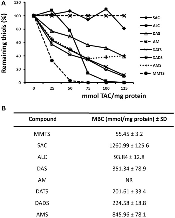
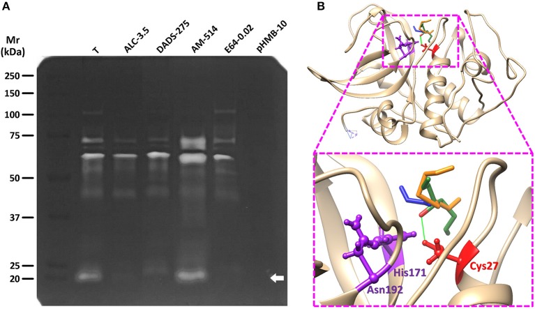
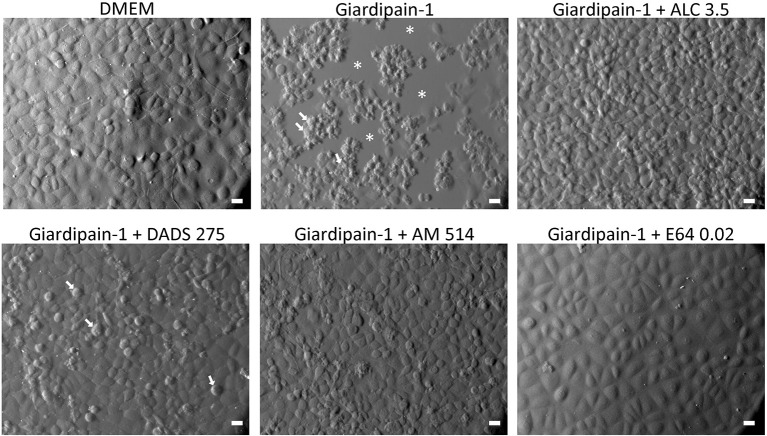
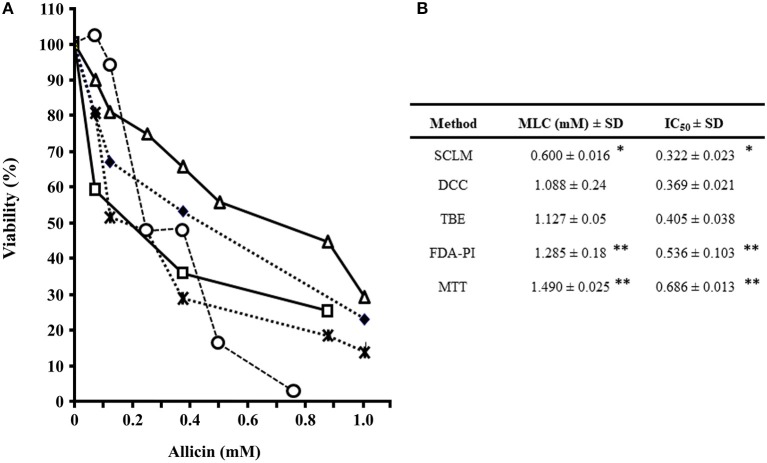
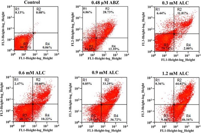
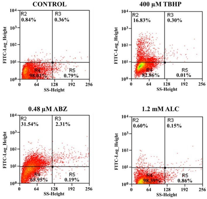
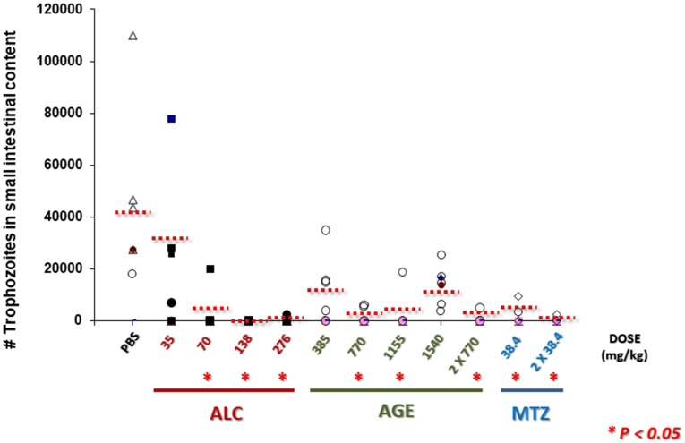
Fresh aqueous extracts (AGEs) and several thioallyl compounds (TACs) from garlic have an important antimicrobial activity that likely involves their interaction with exposed thiol groups at single aminoacids or target proteins. Since these groups are present in Giardia duodenalis trophozoites, in this work we evaluated the anti-giardial activity of AGE and several garlic's TACs. In vitro susceptibility assays showed that AGE affected trophozoite viability initially by a mechanism impairing cell integrity and oxidoreductase activities while diesterase activities were abrogated at higher AGE concentrations. The giardicidal activities of seven TACs were related to the molecular descriptor HOMO (Highest Occupied Molecular Orbital) energy and with their capacity to modify the -SH groups exposed in giardial proteins. Interestingly, the activity of several cysteine proteases in trophozoite lysates was inhibited by representative TACs as well as the cytopathic effect of the virulence factor giardipain-1. Of these, allicin showed the highest anti-giardial activity, the lower HOMO value, the highest thiol-modifying activity and the greatest inhibition of cysteine proteases. Allicin had a cytolytic mechanism in trophozoites with subsequent impairment of diesterase and oxidoreductase activities in a similar way to AGE. In addition, by electron microscopy a marked destruction of plasma membrane and endomembranes was observed in allicin-treated trophozoites while cytoskeletal elements were not affected. In further flow cytometry analyses pro-apoptotic effects of allicin concomitant to partial cell cycle arrest at G2 phase with the absence of oxidative stress were observed. In experimental infections of gerbils, the intragastric administration of AGE or allicin decreased parasite numbers and eliminated trophozoites in experimentally infected animals, respectively. These data suggest a potential use of TACs from garlic against G. duodenalis and in the treatment of giardiasis along with their additional benefits in the host's health.

Keywords: Giardia duodenalis; allicin; garlic; giardiasis; thioallyl compounds; trophozoite damage.


Figures

Figure 1
Figure 1
Evaluation of susceptibility of G. duodenalis trophozoites to aqueous garlic extract (AGE) using four methods. (A) Parasites (1 × 10⁶) were exposed to increasing quantities of 200 μg/mL AGE for 24 h and cellular viability was determined by four methods: subculture in liquid medium (open circles), FDA-PI staining (filled squares), direct cell count (filled diamonds), and MTT reduction (open triangles). Values correspond to results from three independent experiments. (B) The values of the inhibitory concentrations at 50% (IC50) and 100% (MLC) were calculated by the least square method. *Significantly higher than any of the other methods at P < 0.05.
Figure 2
Figure 2
Linear regression model for the MLC-EHOMO relationship of thioallyl compounds (TACs). The values of giardicidal activity (MLC) and the electronic descriptor EHOMO of the six TACs included as “drivers” were processed by multiple linear regression and the value of EHOMO calculated for the “test” compound diallyl trisulfide (DATS) was interpolated in the model (dotted line) to predict the MLC of this compound and to compare it with its experimetal MLC. Abbreviations of compound names are as indicated in section Materials and Methods.
Figure 3
Figure 3
Inhibition of formation of thiol- dithionitrobenzoic acid (DTNB) complexes in protein extracts of G. duodenalis trophozoites by thioallyl compounds (TACs). (A) Parasite extracts (100 μg) were incubated with increasing concentrations of the indicated TACs then the thiol-derivatizing agent DTNB was added to the mixture. Samples without TAC were taken as controls with 100% thiols. Points represent the mean values of two independent experiments. Lines are as follows: S-allyl Cysteine (filled diamonds), Allicin (filled squares), Diallyl Sulfide (open triangles), Allyl Mercaptan (taches), Diallyl Trisulfide (astereisks), Diallyl Disulfide (open circles), Allyl-Methyl Sulfide (crosses), and Methyl Methanethiosulfonate (closed circles). (B) The values of the maximal blocking concentrations (MBC) for each TAC were calculated using a standard curve using cysteine.
Figure 4
Figure 4
Effect of representative TACs on the proteolytic activity of G. duodenalis trophozoite lysates and docking with giardipain-1. (A) Trophozoite lysates (50 μg) were electrophoretically separated in 10% acrylamide-0.2% gelatin gels and cysteine protease activities were developed and detected by DTT treatment at pH 5.5 and Coomassie blue staining. Lanes in which extracts were pre-incubated with the three TACs tested (ALC, DADS, and AM) are indicated along with the concentration values (mM) used in each case. The thiol-active inhibitor pHMB (10 mM) and the cathepsin B-specific inhibitor E64 (20 μM) were included as controls. Molecular weight standards and its relative mobility (Mr) are as indicated at the left. The arrow points to the band containing the cysteine protease giardipain-1. (B) The most favored docking position of representative TACs (in stick conformation, green: ALC, orange: DADS, blue: AM) at the proximity of the catalytic Cys27 of giardipain-1 in which the Oxygen atom of ALC is predicted to form a bridge with the Hydrogen G1 of Cys27 at a distance of 2.425 Å (green line). The three catalytic residues (Cys26, His171, and Asn192) are represented in ball-and-stick conformation.
Figure 5
Figure 5
Effect of garlic TACs on the cytolytic damage caused by giardipain-1 in epithelial IEC6 cell monolayers. Confluent IEC6 cells were exposed for 100 min at 37°C to affinity chromatography-purified giardipain-1 alone or pre-incubated with the concentrations indicated (in mM) of representative TACs that caused inhibition of its protease activity in zymograms and monolayers were observed by Nomarski optics. Asterisks denote zones of monolayer destruction and arrows point to cells undergoing apoptotic death as indicated by membrane blebbing, shrinkage, and shape degeneration. Bars: 20 μm.
Figure 6
Figure 6
Evaluation of suceptibility of G. duodenalis trophozoites to allicin using five methods. (A) Parasites (1 × 10⁶) were exposed to increasing quantities of 2.5% ALC for 24 h and cellular viability was determined by several methods as previously described (Argüello-García et al., 2004). Values correspond to results from three independent experiments. Lines are as follows: subculture in liquid medium (open circles), fluorescein diacetate-propidium iodide staining (FDA-PI) staining (filled diamonds), direct cell count (taches), 3-(4,5-dimethyl-2-thiazolyl)-2,5-diphenyl-2-H-tetrazolium bromide (MTT) reduction (open triangles) and trypan blue exclusion (open squares). (B) The values of IC50 and MLC were calculated by the least square method. *Significantly lower than any of the other methods at P < 0.05. **Significantly higher than SCLM, DCC, and TBE (P < 0.05).
Figure 7
Figure 7
Analysis by scanning (A) and transmission (B) electron microscopy of G. duodenalis trophozoites exposed to allicin. (A) Parasites (1 × 10⁶) were either not exposed (A,B) or exposed to 600 μM ALC (C–F) for 24 h or 1.2 mM ALC for 3 h (G,H) at 37°C. Trophozoites are shown in ventral (A,C, E–G) and dorsal (B,D,H) views. The higher roughness of cell surface in images C–H as compared to A,B, except over the ventral disk, are indicative of a degree of membrane blebbing. Arrows denote the so-called “bared area” of the ventral disk and asterisks point to tip rounding at flagella. (B) Parasites not exposed (1, 3) and exposed to 600 μM ALC (2, 4) for 24 h at 37°C. Trophozoites are shown in longitudinal (1, 2) and cross-sections (3, 4). Arrows indicate the plasma membranes, asterisks show flagellar axonemes and crosses show inner peripheral areas of nucleus with chromatin condensation. N, nuclei; VD, ventral disk. Bars correspond to the indicated size.
Figure 8
Figure 8
Allicin promotes apoptosis-like death in G. duodenalis trophozoites. Parasites (1 × 10⁶) were exposed to 1.0% double distilled water pH 6.5 (Control) or the indicated concentrations of ALC for 24 h at 37°C then stained with annexin V-PI and processed by flow cytometry. Data shown in the figure are as follows: quadrant R1 are cells positive for necrosis (PI+); quadrant R2 are cells positive for late apoptosis (annexin V+/PI+); quadrant R3 are cells negative for both markers and quadrant R4 are cells positive for early apoptosis (annexin V+). As a positive control, trophozoites were exposed to the indicated cytotoxic concentration of albendazole (ABZ). Percentages of total cells are indicated within each quadrant. The dot plots are representative of two independent experiments.
Figure 9
Figure 9
Allicin does not cause reactive oxygen species (ROS) formation in G. duodenalis trophozoites. Parasites (1 × 10⁶) were exposed to 1.0% double distilled water pH 6.5 (Control) or the indicated concentrations of ALC, tert-butyl hydroperoxide (TBHP, positive control) or prooxidant ABZ for 24 h at 37°C then incubated with the ROS-monitoring agent H2DCFDH and processed by flow cytometry. Data shown in the figure are as follows: quadrants R2-R3 are cells positive for ROS and quadrants R4-R5 are cells negative for ROS. Percentages of total cells are indicated within each quadrant. The dot plots are representative of two independent experiments.
Figure 10
Figure 10
Allicin partially arrests G. duodenalis trophozoites at G2 phase. Parasites (1 × 10⁶) were exposed to 1.0% double distilled water pH 6.5 (Control) or the indicated concentrations of ABZ (positive control for cell cycle arrest) or ALC for 24 h at 37°C. Nuclei of permeabilised and RNAse A-treated cells were stained with PI and processed by flow cytometry. The areas under curve (AUC) corresponding to G1, S, G2, or G2/M cell cycle phases are indicated in the control histogram. Percentages of cells within each AUC are shown in the table at the bottom of histograms. Data are representative of two independent experiments.
Figure 11
Figure 11
AGE and allicin diminish and eliminate G. duodenalis trophozoites in small intestines of infected gerbils. Male M. unguiculatus were intragastrically inoculated with 1 × 10⁶ trophozoites per os. At day 10 p.i. gerbils were treated with AGE (single or doubled doses), ALC at single doses or metronidazole (MTZ) at single or double dose as indicated. At day 13 p.i. trophozoites in intestinal contents (10 cm-long sections) were counted. Punctuated lines indicate the mean values of intestinal trophozoites for each group. Asterisks indicate counts significantly lower than control group (PBS) at P < 0.05.

Similar articles

Cited by

References

    1. Adam R. D. (2001). Biology of Giardia lamblia. Clin. Microbiol. Rev. 14, 447–475. 10.1128/CMR.14.3.447-475.2001 - DOI - PMC - PubMed
    1. Ali V., Nozaki T. (2007). Current therapeutics, their problems, and sulfur-containing-amino-acid-metabolism as a novel target against infections by “amitochondriate” protozoan parasites. Clin. Microbiol. Rev. 20, 164–187. 10.1128/CMR.00019-06 - DOI - PMC - PubMed
    1. Alnassan A. A., Thabet A., Daugschies A., Bangoura B. (2015). In vitro efficacy of allicin on chicken Eimeria tenella sporozoites. Parasitol. Res. 114, 3913–3915. 10.1007/s00436-015-4637-2 - DOI - PubMed
    1. Ankarklev J., Jerlström-Hultqvist J., Ringqvist E., Troell K., Svärd S. G. (2010). Behind the smile: cell biology and disease mechanisms of Giardia species. Nat. Rev Microbiol. 8, 413–422. 10.1038/nrmicro2317 - DOI - PubMed
    1. Ankri S., Mirelman D. (1999). Antimicrobial properties of allicin from garlic. Microbes Infect. 1, 125–129. 10.1016/S1286-4579(99)80003-3 - DOI - PubMed

Publication types

MeSH terms