

The Host Response in Tissue Engineering: Crosstalk Between Immune cells and Cell-laden Scaffolds
- PMID: 30374467
- PMCID: PMC6203342
- DOI: 10.1016/j.cobme.2018.03.006
The Host Response in Tissue Engineering: Crosstalk Between Immune cells and Cell-laden Scaffolds
Abstract
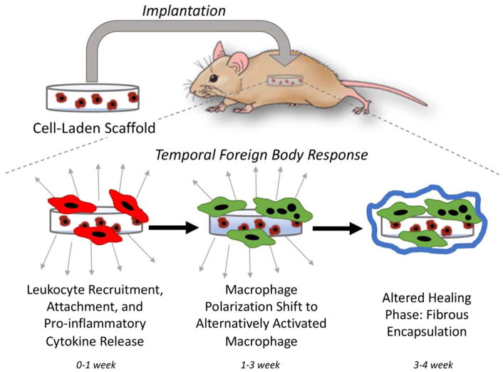
Implantation of cell-laden scaffolds is a promising strategy for regenerating tissue that has been damaged due to injury or disease. However, the act of implantation initiates an acute inflammatory response. If the scaffold is non-biologic (i.e., a modified biologic scaffold or synthetic-based scaffold), inflammation will be prolonged through the foreign body response (FBR), which eventually forms a fibrous capsule and walls off the implant from the surrounding host tissue. This host response, from a cellular perspective, can create a harsh environment leading to long-lasting effects on the tissue engineering outcome. At the same time, cells embedded within the scaffold can respond to this environment and influence the interrogating immune cells (e.g., macrophages). This crosstalk, depending on the type of cell, can dramatically influence the host response. This review provides an overview of the FBR and highlights important and recent advancements in the host response to cell-laden scaffolds with a focus on the impact of the communication between immune cells and cells embedded within a scaffold. Understanding this complex interplay between the immune cells, notably macrophages, and the tissue engineering cells is a critically important component to a successful in vivo tissue engineering therapy.
Keywords: Foreign Body Response; Macrophage; Mesechymal Stem Cells; Scaffold.
Conflict of interest statement
Conflict of interest The authors declare no conflicts of interest.
Figures
Similar articles
-
Immunomodulation by mesenchymal stem cells combats the foreign body response to cell-laden synthetic hydrogels.Biomaterials. 2015 Feb;41:79-88. doi: 10.1016/j.biomaterials.2014.11.020. Epub 2014 Dec 5. Biomaterials. 2015. PMID: 25522967 Free PMC article.
-
Influence of scaffold design on host immune and stem cell responses.Semin Immunol. 2017 Feb;29:62-71. doi: 10.1016/j.smim.2017.03.001. Epub 2017 Apr 26. Semin Immunol. 2017. PMID: 28431919 Review.
-
A Computational Model of Osteochondral Defect Repair Following Implantation of Stem Cell-Laden Multiphase Scaffolds.Tissue Eng Part A. 2017 Jan;23(1-2):30-42. doi: 10.1089/ten.TEA.2016.0175. Epub 2016 Nov 10. Tissue Eng Part A. 2017. PMID: 27712189
-
Host Response and Neo-Tissue Development during Resorption of a Fast Degrading Supramolecular Electrospun Arterial Scaffold.Bioengineering (Basel). 2018 Aug 6;5(3):61. doi: 10.3390/bioengineering5030061. Bioengineering (Basel). 2018. PMID: 30082586 Free PMC article.
-
Insights in the host response towards biomaterial-based scaffolds for cancer therapy.Front Bioeng Biotechnol. 2023 Jun 5;11:1149943. doi: 10.3389/fbioe.2023.1149943. eCollection 2023. Front Bioeng Biotechnol. 2023. PMID: 37342507 Free PMC article. Review.
Cited by
-
Resorbable Biomaterials Used for 3D Scaffolds in Tissue Engineering: A Review.Materials (Basel). 2023 Jun 8;16(12):4267. doi: 10.3390/ma16124267. Materials (Basel). 2023. PMID: 37374451 Free PMC article. Review.
-
Engineering Musculoskeletal Grafts for Multi-Tissue Unit Repair: Lessons From Developmental Biology and Wound Healing.Front Physiol. 2021 Aug 24;12:691954. doi: 10.3389/fphys.2021.691954. eCollection 2021. Front Physiol. 2021. PMID: 34504435 Free PMC article. Review.
-
Protein Based Biomaterials for Therapeutic and Diagnostic Applications.Prog Biomed Eng (Bristol). 2022 Jan;4(1):012003. doi: 10.1088/2516-1091/ac2841. Epub 2021 Oct 26. Prog Biomed Eng (Bristol). 2022. PMID: 34950852 Free PMC article.
-
Efficient Decellularization of the Full-Thickness Rat-Derived Abdominal Wall to Produce Acellular Biologic Scaffolds for Tissue Reconstruction: Promising Evidence Acquired from In Vitro Results.Bioengineering (Basel). 2023 Aug 1;10(8):913. doi: 10.3390/bioengineering10080913. Bioengineering (Basel). 2023. PMID: 37627798 Free PMC article.
-
Mesenchymal stem cell-inspired microgel scaffolds to control macrophage polarization.Bioeng Transl Med. 2021 Mar 21;6(2):e10217. doi: 10.1002/btm2.10217. eCollection 2021 May. Bioeng Transl Med. 2021. PMID: 34027099 Free PMC article.
References
-
- Ratner BD, Bryant SJ. Biomaterials: Where we have been and where we are going. Annu Rev Biomed Eng. 2004;6:41–75. - PubMed
-
- Anderson JM. Biological responses to materials. Annu Rev Mater Res. 2001;31:81–110.
Grants and funding
LinkOut - more resources
Full Text Sources
Other Literature Sources