Review
. 2018 Nov;19(Suppl 1):3-14.
doi: 10.1007/s40257-018-0367-4.

Noninvasive Imaging Tools in the Diagnosis and Treatment of Skin Cancers

Affiliations
Review

Noninvasive Imaging Tools in the Diagnosis and Treatment of Skin Cancers

Renato Marchiori Bakos et al. Am J Clin Dermatol. 2018 Nov.

Abstract

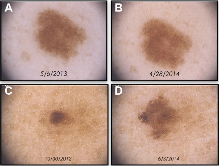
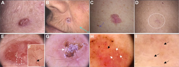
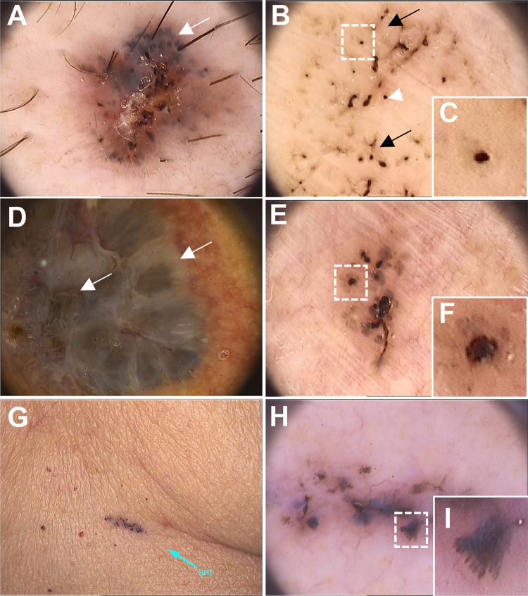
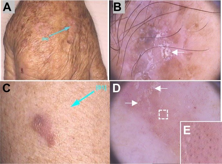
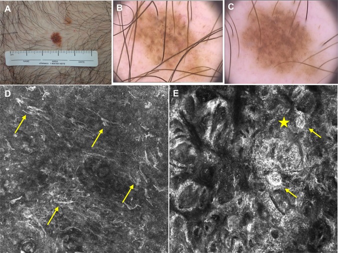
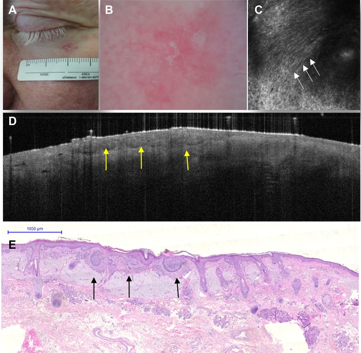
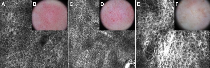
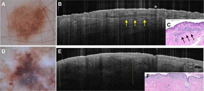
In the 1980s, the increasing incidence of skin cancers prompted the development of noninvasive medical devices to improve skin cancer diagnosis in daily dermatology practice. As a result of the development of these noninvasive techniques, diagnosis is now established earlier and with better accuracy. These advances are of great benefit to high-risk patients, who previously would have had to undergo several excisions. In this review, we focus on the classic technique of dermoscopy and the more recent digital version, as well as on advanced noninvasive imaging techniques, such as reflectance confocal microscopy and optical coherence tomography. On the basis of their specific features, these noninvasive medical devices can be used not only to diagnose and monitor melanoma and nonmelanoma skin cancers but also to choose the best therapy and follow the patient's response to treatment in vivo.


Conflict of interest statement

Conflict of interest

Renato Marchiori Bakos, Tatiana Pinto Blumetti, Rodrigo Roldán-Marín, and Gabriel Salerni declare that they have no conflicts of interest that might be relevant to the content of this manuscript.

Disclosure statement

This article is published as part of a journal supplement wholly funded by Laboratoires dermatologiques Avène.

Figures

Fig. 1
Fig. 1
Follow-up of benign and malignant pigmented lesions by dermoscopy. Stability of a benign lesion over time (a, b). Change of a pigmented lesion over time suggesting malignancy (c, d). Dermoscopy original magnification ×20
Fig. 2
Fig. 2
Facial nonpigmented basal cell carcinoma lesions. Photographs of nodular (a, b), superficial (c), and infiltrative (d) basal cell carcinoma lesions and the corresponding dermoscopic images (original magnification × 20, Fotofinder systems): thick arborizing telangiectasias branching over the lesion (e) and at high magnification (black arrow) (f), and ulceration (white arrow) (g). Microerosions (white arrows) and super fine, short telangiectasias (black arrow) (h). Fine and scattered telangiectasias (black arrows) (i)
Fig. 3
Fig. 3
Dermoscopic features of pigmented basal cell carcinoma lesions. Dermoscopic images (original magnification ×20, Fotofinder systems) showing ulceration and multiple gray dots (white arrows) (a); maple-leaf area (black arrows), spoke-wheel-like area, concentric structures (white arrowheads) (b, high magnification in c); ovoid nests (white arrows) (d); spoke-wheel-like area (e, high magnification in f). Photograph of a small pigmented lesion and angiomas (g) and the corresponding dermoscopic image (h) showing a maple-leaf-like area (high magnification in i)
Fig. 4
Fig. 4
Nonfacial actinic keratosis and Bowen’s disease (adapted from Reinehr et al. [28]). Photographs of nonfacial pigmented actinic keratosis lesions on the dorsa of the hand (a) and the corresponding dermoscopic image (original magnification × 20) with opaque white scales (arrows) and homogenous brown pigmentation (b) (from Reinehr et al. [28]). Bowen’s disease photograph (c) and dermoscopic image showing yellowish scales (arrows) (d) and glomerular vessels at high magnification (e) (Renato Bakos; personal images)
Fig. 5
Fig. 5
Refinement of the diagnosis of ‘featureless melanoma’ by RCM. Clinical (a) and dermoscopic images [first examination (b) and after 6 months of follow-up (c)] of a melanocytic lesion on a patient with atypical nevi syndrome with no previous history of melanoma. The lesion presented a slight change in size and color over 6 months, but showed no specific melanoma features on dermoscopy. In the epidermis, the RCM image (d) (individual image, 0.5 × 0.5 mm) shows an atypical honeycomb pattern and widespread pagetoid infiltration of dendritic atypical cells (arrows). At the dermal–epithelial junction, the RCM image (e) (1.0 × 1.0 mm) shows an atypical meshwork pattern, clusters of dendritic atypical cells (star) at the papillae, and atypical junctional nests (arrows). The histopathologic analysis resulted in diagnosis of a superficial spreading melanoma (Breslow 0.25 mm). Images kindly provided by Dr. Juliana Tavoloni Braga (AC Camargo Cancer Center). RCM reflectance confocal microscopy
Fig. 6
Fig. 6
Refinement of the diagnosis of a superficial basal cell carcinoma lesion with confocal microscopy and optical coherence tomography. Clinical image of an erythematosquamous lesion on the lower eyelid (a). The dermoscopic image (b) shows erythema and scaling, and the confocal image (c) (individual image; 0.5 × 0.5 mm) reveals a ‘streaming pattern’ of cells (elongated keratinocytes following the same axis) in the epidermis (white arrows). The optical coherence tomography image (d) shows epidermal hyporeflective thickening with dark borders and the correspondence to histopathology (hematoxylin–eosin stain) (e) revealing basaloid nests attached to the epidermis (yellow arrows). Images kindly provided by Dr. Elimar Gomes (AC Camargo Cancer Center)
Fig. 7
Fig. 7
Refinement of the diagnosis of keratinocytic lesions with confocal microscopy. AK on reflectance confocal microscopy (a, c, e) and dermoscopy (b, d, f). Dermoscopy reveals patterns of AK of grades I (b), II (d), and III (e) showing a ‘strawberry’ pattern characterized by a red pseudonetwork in b, increasing erythema and scaling in d, and a ‘starbust’ pattern in f. Confocal microscopy (epidermis, 0.5 × 0.5 mm) demonstrates the progress of atypia of the honeycomb pattern, showing keratinocytes with different shapes and sizes increasing the level of atypia within the granular and spinous layer with evolution of the dermoscopy pattern of the AKs. AK actinic keratosis
Fig. 8
Fig. 8
Dermoscopy and optical coherence tomography images of melanocytic lesions: compound dysplastic nevus and melanoma. Dermoscopy image a of a dysplastic nevus showing a 0.5 mm melanocytic lesion with an atypical network pattern. On optical coherence tomography (b), a well-demarcated hyporeflective band surrounded by normal collagen can be visualized. This structure corresponds to an intradermal melanocytic nest on histopathology (c, hematoxylin–eosin stain). The dermoscopy image d of a superficial spreading melanoma (Breslow thickness 0.45 mm). The optical coherence tomography image e shows a focal loss of visualization of collagen in the dermis (shadows illustrated between yellow lines) and histopathology (f, ×10 magnification)

References

    1. Pehamberger H, Steiner A, Wolff K. In vivo epiluminescence microscopy of pigmented skin lesions. I. Pattern analysis of pigmented skin lesions. J Am Acad Dermatol. 1987;17(4):571–583. doi: 10.1016/S0190-9622(87)70239-4. - DOI - PubMed
    1. Marghoob AA, Korzenko AJ, Changchien L, Scope A, Braun RP, Rabinovitz H. The beauty and the beast sign in dermoscopy. Dermatol Surg. 2007;33(11):1388–1391. - PubMed
    1. Kittler H, Pehamberger H, Wolff K, Binder M. Diagnostic accuracy of dermoscopy. Lancet Oncol. 2002;3(3):159–165. doi: 10.1016/S1470-2045(02)00679-4. - DOI - PubMed
    1. Vestergaard ME, Macaskill P, Holt PE, Menzies SW. Dermoscopy compared with naked eye examination for the diagnosis of primary melanoma: a meta-analysis of studies performed in a clinical setting. Br J Dermatol. 2008;159(3):669–676. - PubMed
    1. Huerta-Brogeras M, Olmos O, Borbujo J, Hernandez-Nunez A, Castano E, Romero-Mate A, et al. Validation of dermoscopy as a real-time noninvasive diagnostic imaging technique for actinic keratosis. Arch Dermatol. 2012;148(10):1159–1164. doi: 10.1001/archdermatol.2012.1060. - DOI - PubMed

MeSH terms