

Translation of Carbon-13 EPI for hyperpolarized MR molecular imaging of prostate and brain cancer patients
- PMID: 30375043
- PMCID: PMC6372313
- DOI: 10.1002/mrm.27549
Translation of Carbon-13 EPI for hyperpolarized MR molecular imaging of prostate and brain cancer patients
Abstract
Purpose: To develop and translate a metabolite-specific imaging sequence using a symmetric echo planar readout for clinical hyperpolarized (HP) Carbon-13 (13 C) applications.
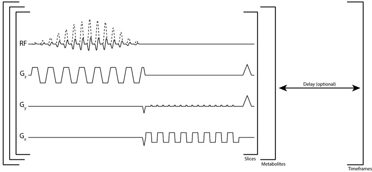
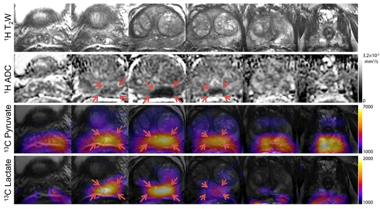
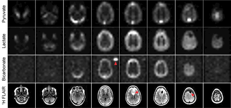
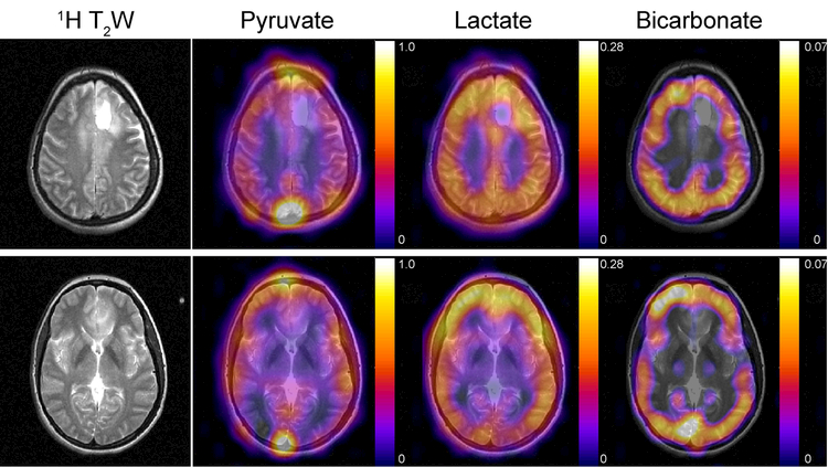
Methods: Initial data were acquired from patients with prostate cancer (N = 3) and high-grade brain tumors (N = 3) on a 3T scanner. Samples of [1-13 C]pyruvate were polarized for at least 2 h using a 5T SPINlab system operating at 0.8 K. Following injection of the HP substrate, pyruvate, lactate, and bicarbonate (for brain studies) were sequentially excited with a singleband spectral-spatial RF pulse and signal was rapidly encoded with a single-shot echo planar readout on a slice-by-slice basis. Data were acquired dynamically with a temporal resolution of 2 s for prostate studies and 3 s for brain studies.
Results: High pyruvate signal was seen throughout the prostate and brain, with conversion to lactate being shown across studies, whereas bicarbonate production was also detected in the brain. No Nyquist ghost artifacts or obvious geometric distortion from the echo planar readout were observed. The average error in center frequency was 1.2 ± 17.0 and 4.5 ± 1.4 Hz for prostate and brain studies, respectively, below the threshold for spatial shift because of bulk off-resonance.
Conclusion: This study demonstrated the feasibility of symmetric EPI to acquire HP 13 C metabolite maps in a clinical setting. As an advance over prior single-slice dynamic or single time point volumetric spectroscopic imaging approaches, this metabolite-specific EPI acquisition provided robust whole-organ coverage for brain and prostate studies while retaining high SNR, spatial resolution, and dynamic temporal resolution.
Keywords: DNP; EPI; hyperpolarization; oncology; pyruvate.
© 2018 International Society for Magnetic Resonance in Medicine.
Figures
References
-
- Kurhanewicz J, Vigneron DB, Brindle K, Chekmenev EY, Comment A, Cunningham CH, DeBerardinis RJ, Green GG, Leach MO, Rajan SS, Rizi RR, Ross BD, Warren WS, Malloy CR. Analysis of Cancer Metabolism by Imaging Hyperpolarized Nuclei: Prospects for Translation to Clinical Research. Neoplasia 2011;13(2):81–97. - PMC - PubMed
-
- Albers MJ, Bok R, Chen AP, Cunningham CH, Zierhut ML, Zhang VY, Kohler SJ, Tropp J, Hurd RE, Yen Y-F, Nelson SJ, Vigneron DB, Kurhanewicz J. Hyperpolarized 13C Lactate, Pyruvate, and Alanine: Noninvasive Biomarkers for Prostate Cancer Detection and Grading. Cancer Res 2008;68(20):8607–8615. - PMC - PubMed
-
- Golman K, Zandt Rit, Lerche M, Pehrson R, Ardenkjaer-Larsen JH. Metabolic Imaging by Hyperpolarized 13C Magnetic Resonance Imaging for In vivo Tumor Diagnosis. Cancer Research 2006;66(22):10855–10860. - PubMed
-
- Day SE, Kettunen MI, Gallagher FA, Hu DE, Lerche M, Wolber J, Golman K, Ardenkjaer-Larsen JH, Brindle KM. Detecting tumor response to treatment using hyperpolarized 13C magnetic resonance imaging and spectroscopy. Nat Med 2007;13(11):1382–1387. - PubMed
Publication types
MeSH terms
Substances
Grants and funding
- P41 EB013598/EB/NIBIB NIH HHS/United States
- R01CA183071/NH/NIH HHS/United States
- P01 CA118816/CA/NCI NIH HHS/United States
- R01EB017449/NH/NIH HHS/United States
- R01 CA183071/CA/NCI NIH HHS/United States
- R01 EB017449/EB/NIBIB NIH HHS/United States
- R01 CA211150/CA/NCI NIH HHS/United States
- UCSF/International
- P01CA118816/NH/NIH HHS/United States
- R01EB017449, R01CA183071, P41EB013598, P01CA118816, and R01CA211150/NH/NIH HHS/United States
- R01CA211150/NH/NIH HHS/United States
- P41EB013598/NH/NIH HHS/United States
LinkOut - more resources
Full Text Sources
Other Literature Sources
Medical
Research Materials
Miscellaneous
