. 2019 Aug;26(8):1428-1441.
doi: 10.1038/s41418-018-0219-z. Epub 2018 Oct 30.

PINK1-dependent mitophagy is driven by the UPS and can occur independently of LC3 conversion

Affiliations

PINK1-dependent mitophagy is driven by the UPS and can occur independently of LC3 conversion

Aleksandar Rakovic et al. Cell Death Differ. 2019 Aug.

Abstract

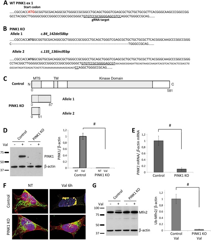
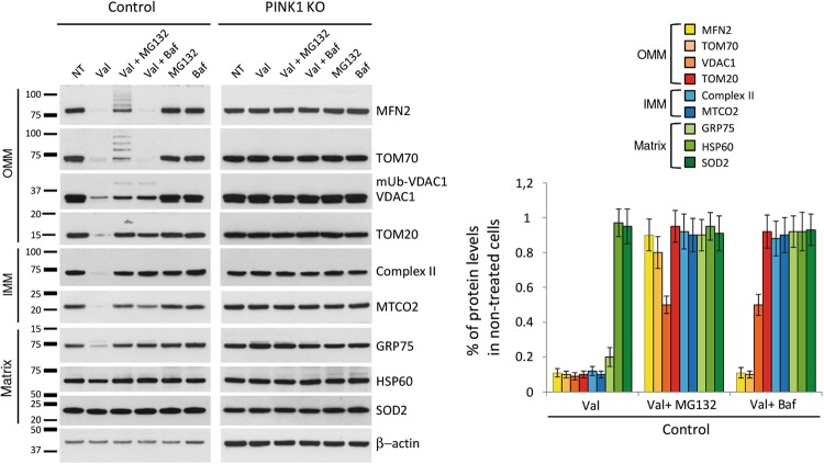
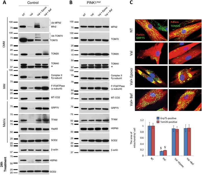
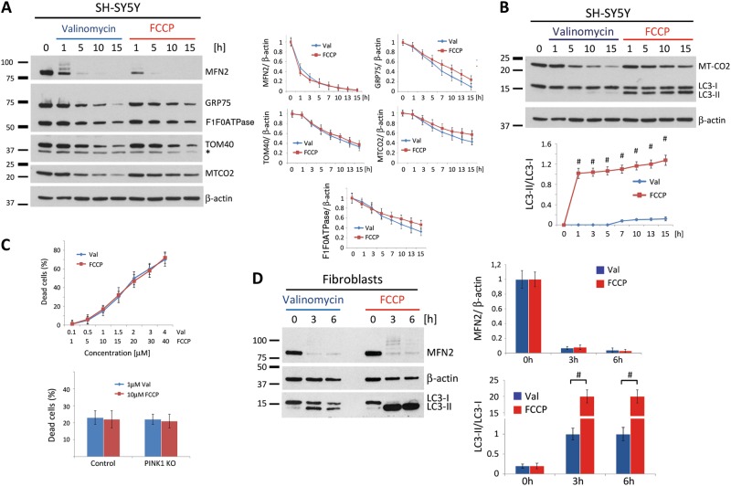
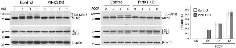
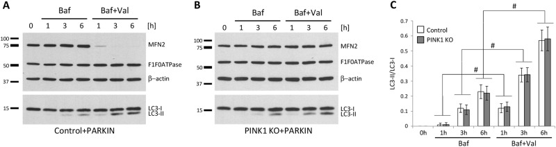
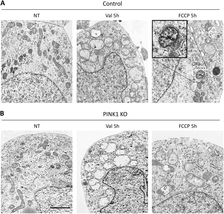
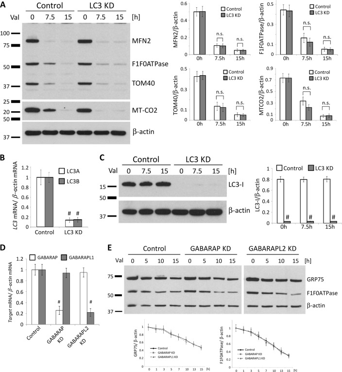
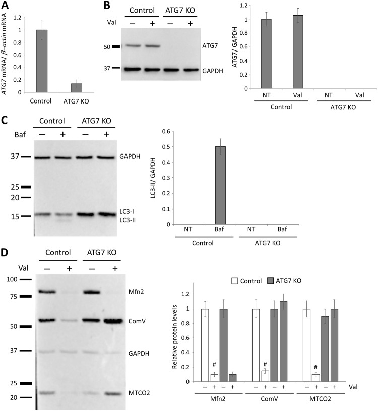
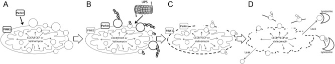
The Parkinson's disease (PD)-related ubiquitin ligase Parkin and mitochondrial kinase PINK1 function together in the clearance of damaged mitochondria. Upon mitochondrial depolarization, Parkin translocates to mitochondria in a PINK1-dependent manner to ubiquitinate outer mitochondrial membrane proteins. According to the current model, the ubiquitin- and LC3-binding adaptor protein SQSTM1 is recruited to mitochondria, followed by their selective degradation through autophagy (mitophagy). However, the role of the ubiquitin proteasome system (UPS), although essential for this process, still remains largely elusive. Here, we investigated the role of the UPS and autophagy by applying the potassium ionophore Valinomycin in PINK1-deficient human fibroblasts and isogenic neuroblastoma cell lines generated by CRISPR/Cas9. Although identical to the commonly used CCCP/FCCP in terms of dissipating the mitochondrial membrane potential and triggering complete removal of mitochondria, Valinomycin did not induce conversion of LC3 to its autophagy-related form. Moreover, FCCP-induced conversion of LC3 occurred even in mitophagy-incompetent, PINK1-deficient cell lines. While both stressors required a functional UPS, the removal of depolarized mitochondria persisted in cells depleted of LC3A and LC3B. Our study highlights the importance of the UPS in PINK1-/Parkin-mediated mitochondrial quality control. In contrast, activation of autophagy, monitored through conversion of LC3, is likely induced by depolarizing-agent-induced toxicity in a PINK1-/Parkin-independent manner.


Conflict of interest statement

The authors declare that they have no conflict of interest.

Figures

Fig. 1
Fig. 1
CRISPR/Cas9-mediated knockout of PINK1 in neuroblastoma cells. a Neuroblastoma (SH-SY5Y) cells were transfected with episomal vectors expressing Cas9 and gRNA targeting the underlined sequence located in exon 1 of the PINK1 gene. b The PINK1 knockout (PINK1KO) clonal cell line carries compound-heterozygous mutations in PINK1 ([c.84_142del58bp]+[c.135_136ins95bp]). c Schematic representation of the wildtype PINK1 protein and putative truncated forms of the PINK1 protein in PINK1KO cells. Areas shaded in gray represent a frame-shifted protein. d To detect endogenous PINK1 protein, control and PINK1KO cells were treated with Valinomycin for 6 h and analyzed by western blotting using antibodies against PINK1. Protein levels of PINK1 were quantified and normalized to levels of β-actin. e PINK1 mRNA expression in control and PINK1KO SH-SY5Y cells. The values represent means ± SD from three independent measurements. f Control and PINK1KO cells treated with Valinomycin for 6 h were fixed and immunostained using antibodies against Parkin (red) and the mitochondrial matrix protein GRP75 (green). g Immunoblot of untreated and Valinomycin-treated control and PINK1KO cells probed with an antibody against MFN2. Levels of ubiquitinated MFN2 (Ub-MFN2) in Valinomycin-treated cells were normalized to levels of β-actin. #p < 0.01
Fig. 2
Fig. 2
Inhibition of the UPS or lysosomal system prevents removal of depolarized mitochondria in neuroblastoma cells. Control or PINK1KO neuroblastoma cells engineered to stably overexpress Parkin were treated with Valinomycin alone or in combination with MG132 or Bafilomycin A1 for 16 h. In addition, cells were treated with either MG132 or Bafilomycin A1. Cells were harvested and analyzed by western blot using antibodies against mitochondrial proteins localized in different mitochondrial compartments (outer mitochondrial membrane (OMM), inner mitochondrial membrane (IMM), and matrix). Protein levels in control neuroblastoma cells were quantified and normalized to levels of β-actin. Values represent means ± SD from three independent measurements.
Fig. 3
Fig. 3
Inhibition of the UPS or lysosomal system prevents removal of depolarized mitochondria in human fibroblasts. a Control and b PINK1mut fibroblasts stably overexpressing Parkin were treated with Valinomycin only or in combination either with Epoxomicin or with Bafilomycin A1 for 16 h. Cells were immunoblotted using antibodies against mitochondrial proteins localized in the three different mitochondrial compartments. β-actin served as loading control. Results with longer (24 h) Valinomycin treatment are also shown for HSP60 and SOD2. *: non-specific band. c Control fibroblasts stably expressing Parkin were treated with Valinomycin only or in combination either with Epoxomicin or with Bafilomycin A1 for 16 h. Cells were immunostained using antibodies against Parkin (red) together with either GRP75 (green) or TOM20 (green). Average area of either GRP75-positive or TOM20-positive mitochondria per cell. Values represent means ± SD from three independent measurements. §p < 0.01 vs. non-treated cells
Fig. 4
Fig. 4
Valinomycin-induced clearance of mitochondria does not require activation of autophagy. Neuroblastoma cells stably overexpressing Parkin were harvested at different time points and analyzed by western blotting using antibodies against a proteins localized in different mitochondrial compartments or against b the autophagy marker LC3. Protein levels were quantified and normalized to levels of β-actin. c Trypan blue exclusion assay test in control neuroblastoma cells overexpressing Parkin treated with various concentration of Valinomycin or FCCP for 16 h (upper panel). Trypan blue exclusion assay in control and PINK1KO neuroblastoma cells overexpressing Parkin treated with either Valinomycin or FCCP for 16 h (lower panel). d Human dermal fibroblasts from a healthy control overexpressing Parkin were treated with either Valinomycin or FCCP. Cells were harvested 7.5 and 15 h upon Valinomycin treatment and immunoblotted using antibodies against MFN2 and LC3. Values represent means ± SD from three independent measurements. #p < 0.01 vs. Valinomycin. Asterisk: nonspecific band
Fig. 5
Fig. 5
FCCP initiates LC3 conversion in mitophagy-incompetent cells. Control and PINK1KO neuroblastoma cells expressing endogenous levels of Parkin were treated with either Valinomycin or FCCP for 6 h and harvested at different time points. Whole cell lysates were analyzed by western blotting using antibodies against MFN2 or LC3. Protein levels of LC3 were quantified and normalized to levels of β-actin. Values represent means ± SD from three independent measurements
Fig. 6
Fig. 6
Autophagy flux is comparable between Valinomycin-treated control and mitophagy-incompetent PINK1 knockout cells. a Control and b PINK1KO neuroblastoma cells overexpressing Parkin were treated with Bafilomycin A1 alone or in combination with Valinomycin for 6 h and harvested at different time points. Whole cell lysates were analyzed by western blotting using antibodies against MFN2, the α subunit of F1F0ATPase or LC3. c Protein levels of LC3 were quantified and normalized to levels of β-actin. Values represent means ± SD from three independent measurements.
Fig. 7
Fig. 7
Valinomycin does not induce the formation of autophagic structures. a Control and b PINK1KO neuroblastoma cells stably overexpressing Parkin were treated with either Valinomycin or FCCP for 5 h and compared to non-treated cells. Cells were fixed and prepared for electron microscopy. Treatment with Valinomycin induced mitochondrial swelling and accumulation in the perinuclear region in both controls and PINK1KO cells. Importantly, no isolation membranes or any other autophagic structures were observed. In contrast, upon FCCP treatment, some mitochondria were surrounded by autophagosome-like structures (inset) only in control but not in PINK1KO cells. Val Valinomycin, NT non-treated; scale bar: 2 μm
Fig. 8
Fig. 8
LC3, GABARAP, or GABARAPL1 are not required for Valinomycin-induced removal of mitochondria. Neuroblastoma cells (SH-SY5Y) stably overexpressing Parkin were transduced with lentiviral particles expressing scrambled shRNA (Control) or shRNAs against both LC3A and LC3B (LC3KD). a Control and LC3KD cells were treated with Valinomycin for 15 h and harvested at different time points. Whole cell lysates were analyzed by western blotting using antibodies against different mitochondrial proteins. Protein levels of mitochondrial proteins were quantified and normalized to levels of β-actin. The knockdown efficiency of LC3 was confirmed b by real-time PCR and c by western blotting. d GABARAP or GABARAPL2 knockdown efficiency in SH-SY5Y overexpressing Parkin was determined by real-time PCR. e Control, GABARAPKD, or GABARAPL1KD cells were treated with Valinomycin for 15 h and harvested at different time points. Whole cell lysates were analyzed by western blotting using antibodies against mitochondrial proteins GRP75 and α subunit of F1F0ATPase. Protein levels of mitochondrial proteins were quantified and normalized to levels of β-actin. Values represent means ± SD from three independent measurements. #p < 0.01 vs. control. Val Valinomycin, n.s. not significant
Fig. 9
Fig. 9
Autophagy is not involved in the removal of outer mitochondrial membrane proteins. a ATG7 mRNA expression in control and ATG7 knockout neuroblastoma cells (ATG7KO). b Protein levels of ATG7 in control and ATG7KO levels. c Lack of the Bafilomycin A-induced conversion of non-autophagic LC3-I to autophagic LC3-II confirmed impaired autophagic flux in ATG7KO cells. d Control and ATG7KO 7 cells stably expressing Parkin were treated with Valinomycin for 15 h. Whole cell lysates of non-treated and Valinomycin-treated cells were analyzed by western blotting using antibodies against the outer mitochondrial membrane protein MFN2 and the inner mitochondrial membrane proteins α subunit of F1F0ATPase (ComV) and MTCO2. Protein levels of mitochondrial proteins were quantified and normalized to levels of GAPDH. Values represent means ± SD from three independent measurements. #p < 0.01 vs. control. Val Valinomycin
Fig. 10
Fig. 10
Putative model of PINK1-/Parkin-dependent removal of depolarized mitochondria. a Valinomycin (or CCCP/FCCP)-mediated mitochondrial depolarization initiates mitochondrial accumulation of PINK1 and mitochondrial translocation of Parkin. In parallel, both Valinomycin and CCCP/FCCP increase intramitochondrial pressure. b Parkin-mediated ubiquitination of outer mitochondrial membrane (OMM) proteins and UPS-mediated degradation of (large) OMM proteins. c UPS-mediated proteolysis of large OMM, increased intramitochondrial pressure and intrinsic pressure of IMM on the OMM leads to its destabilization and rupture. d Prolonged Valinomycin- or CCCP/FCCP-induced increase of the intramitochondrial pressure and lack of OMM destabilize the IMM and lead to its rupture. Mitochondrial debris is removed by lysosome-mediated proteolysis and lipolysis

Similar articles

Cited by

References

    1. Kasten M, et al. Genotype–phenotype relations for the Parkinson’s disease genes Parkin, PINK1, DJ1: MDSGene systematic review. Mov Disord. 2018;33:730–41. doi: 10.1002/mds.27352. - DOI - PubMed
    1. Clark IE, et al. Drosophila PINK1 is required for mitochondrial function and interacts genetically with Parkin. Nature. 2006;441:1162–6. doi: 10.1038/nature04779. - DOI - PubMed
    1. Exner N, et al. Loss-of-function of human PINK1 results in mitochondrial pathology and can be rescued by Parkin. J Neurosci. 2007;27:12413–8. doi: 10.1523/JNEUROSCI.0719-07.2007. - DOI - PMC - PubMed
    1. Park J, et al. Mitochondrial dysfunction in Drosophila PINK1 mutants is complemented by Parkin. Nature. 2006;441:1157–61. doi: 10.1038/nature04788. - DOI - PubMed
    1. Rakovic A, et al. Phosphatase and tensin homolog (PTEN)-induced putative kinase 1 (PINK1)-dependent ubiquitination of endogenous Parkin attenuates mitophagy: study in human primary fibroblasts and induced pluripotent stem cell-derived neurons. J Biol Chem. 2013;288:2223–37. doi: 10.1074/jbc.M112.391680. - DOI - PMC - PubMed

Publication types

MeSH terms