Review
. 2019 Jan;17(1):6-18.
doi: 10.1111/jth.14322. Epub 2018 Dec 13.

Exocytosis of Weibel-Palade bodies: how to unpack a vascular emergency kit

Affiliations
Review

Exocytosis of Weibel-Palade bodies: how to unpack a vascular emergency kit

M Schillemans et al. J Thromb Haemost. 2019 Jan.

Abstract

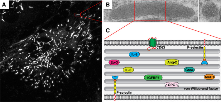
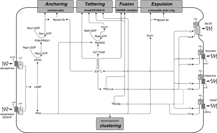
The blood vessel wall has a number of self-healing properties, enabling it to minimize blood loss and prevent or overcome infections in the event of vascular trauma. Endothelial cells prepackage a cocktail of hemostatic, inflammatory and angiogenic mediators in their unique secretory organelles, the Weibel-Palade bodies (WPBs), which can be immediately released on demand. Secretion of their contents into the vascular lumen through a process called exocytosis enables the endothelium to actively participate in the arrest of bleeding and to slow down and direct leukocytes to areas of inflammation. Owing to their remarkable elongated morphology and their secretory contents, which span the entire size spectrum of small chemokines all the way up to ultralarge von Willebrand factor multimers, WPBs constitute an ideal model system for studying the molecular mechanisms of secretory organelle biogenesis, exocytosis, and content expulsion. Recent studies have now shown that, during exocytosis, WPBs can undergo several distinct modes of fusion, and can utilize fundamentally different mechanisms to expel their contents. In this article, we discuss recent advances in our understanding of the composition of the WPB exocytotic machinery and how, because of its configuration, it is able to support WPB release in its various forms.

Keywords: Weibel-Palade bodies; endothelial cells; exocytosis; von Willebrand disease; von Willebrand factor.


Conflict of interest statement

The authors state that they have no conflict of interest.

Figures

Figure 1
Figure 1
Weibel–Palade bodies (WPBs), secretory organelles of the endothelium. (A) Endothelial cells containing characteristic elongated WPBs visualized by von Willebrand factor (VWF) immunostaining. (B) WPB ultrastructure, with a longitudinal section (left) showing internal striations, and a cross‐section (right) showing bundles, which represent densely packed VWF tubules. (C) Cartoon representation of WPB cargo. Ang‐2, angiopoietin‐2; Eo‐3, eotaxin‐3; GROα, growth regulated oncogene α; IGFBP7, insulin‐like growth factor‐binding protein 7; IL‐6, interleukin‐6; IL‐8, interleukin‐8; MCP‐1, monocyte chemoattractant protein‐1; OPG, osteoprotegerin.
Figure 2
Figure 2
The endothelial secretory pathway. von Willebrand factor (VWF) secretion occurs via three pathways: (i) constitutive secretion of low molecular weight VWF, which is primarily released at the basolateral side of the endothelium; and (ii) basal and (iii) regulated secretion of high molecular weight VWF from Weibel–Palade bodies (WPBs), which is primarily directed towards the apical surface. From the large number of WPBs that undergo exocytosis upon stimulated release, ultralarge VWF (UL‐VWF) multimers emerge that assemble into VWF strings on the apical side of the endothelium.
Figure 3
Figure 3
Signaling cascades in Weibel–Palade body (WPB) exocytosis. Ca2+‐mediated and cAMP‐mediated secretagogues that trigger WPB exocytosis use distinct and common signaling circuits that converge at effector pathways that control anchoring, tethering, vesicle fusion, and actin contractility. AC, adenylyl cyclase; ATP, adenosine triphosphate; CaM, calmodulin; [Ca2+]i, intracellular free Ca2+ concentration; DAG, diacylglycerol; EPAC, exchange protein that is directly activated by cAMP; Gb3, ceramide trihexoside; H1R, histamine H1 receptor; IP3, inositol 1,4,5‐triphosphate; PAR1, protease‐activated receptor 1; PI3K, phosphatidylinositol 3‐kinase; PIPa, phosphatidylinositol 4,5‐bisphosphate; PIP2, phosphatidylinositol 4,5‐bisphosphate; PKA, protein kinase A; PKC, protein kinase C; PLC, phospholipase C; PLD1, phospholipase D1; SNARE, soluble N‐ethylmaleimide‐sensitive factor attachment protein receptor; Stx1B, Shiga toxin 1B; V2F, vasopressin‐2 receptor; VEGF, vascular endothelial growth factor; VEGFR2, vascular endothelial growth factor receptor 2; β2‐AR, β2‐adrenergic receptor.
Figure 4
Figure 4
The Weibel–Palade body (WPB) exocytotic machinery. Rab effector complexes mediate anchoring of WPBs to the cytoskeleton, tethering to the plasma membrane, and interactions with the soluble N‐ethylmaleimide‐sensitive factor attachment protein receptor (SNARE) fusion machinery. The Rab27A–MyRIP–myosin Va complex anchors WPBs to the actin cytoskeleton. Munc13‐4 can be recruited by Rab GTPase‐dependent (Rab15/Rab27A) or by Rab‐independent mechanisms, and tethers WPBs to membrane fusion sites via the annexin A2–S100A10 complex. The Rab27A–Slp4‐a complex docks WPBs and forms the link between the WPB and the SNARE complex via members of the syntaxin‐binding protein (STXBP) family. VAMP, vesicle‐associated membrane protein.
Figure 5
Figure 5
Secretory modes of Weibel–Palade body (WPB) exocytosis. (A) Cartoon of a WPB with WPB‐localized v‐SNAREs (blue) engaging with target membrane‐localized t‐SNAREs (pink and green), thereby bringing together donor and acceptor membranes. Top: a narrow fusion pore is formed that permits release of small cargo. Lingering‐kiss fusion resulting from premature closure of the fusion pore results in large cargo (von Willebrand factor [VWF]) being retained in a collapsed granule, whereas expansion of the fusion pore results in full fusion followed by explosive release of ultralarge VWF strings. Top right: compound fusion of WPBs, possibly including lingering‐kiss end‐products, leads to the formation of an enlarged secretory pod that eventually undergoes fusion at the plasma membrane. Bottom left: cumulative or sequential fusion, in which a primary granule undergoes fusion at the plasma membrane, which is followed by secondary fusion events in the postfusion WPB, ultimately leading to extensive, ginger root‐like fusion structures. (B) Two models of SNARE‐mediated cumulative fusion. (Bi) Sequential fusion events are supported by cognate SNARE assemblies on individual, prefusion WPBs. (Bii) Cumulative release is supported by rapid membrane mixing, recruiting plasma membrane SNARE components into the postfusion WPB. SNARE, soluble N‐ethylmaleimide‐sensitive factor attachment protein receptor; VAMP, vesicle‐associated membrane protein.

Similar articles

Cited by

References

    1. Weibel ER, Palade GE. New cytoplasmic components in arterial endothelia. J Cell Biol 1964; 23: 101–12. - PMC - PubMed
    1. Ruggeri ZM. Von Willebrand factor, platelets and endothelial cell interactions. J Thromb Haemost 2003; 1: 1335–42. - PubMed
    1. Valentijn KM, Sadler JE, Valentijn JA, Voorberg J, Eikenboom J. Functional architecture of Weibel–Palade bodies. Blood 2011; 117: 5033–43. - PMC - PubMed
    1. Springer TA. von Willebrand factor, Jedi knight of the bloodstream. Blood 2014; 124: 1412–26. - PMC - PubMed
    1. Leebeek FWG, Eikenboom JCJ. Von Willebrand's disease. N Engl J Med 2016; 375: 2067–80. - PubMed

Publication types

Substances