. 2018 Nov 14;140(45):15261-15269.
doi: 10.1021/jacs.8b07988. Epub 2018 Oct 30.

Use of a Fluorescent Analogue of a HBV Core Protein-Directed Drug To Interrogate an Antiviral Mechanism

Affiliations

Use of a Fluorescent Analogue of a HBV Core Protein-Directed Drug To Interrogate an Antiviral Mechanism

Smita Nair et al. J Am Chem Soc. .

Abstract

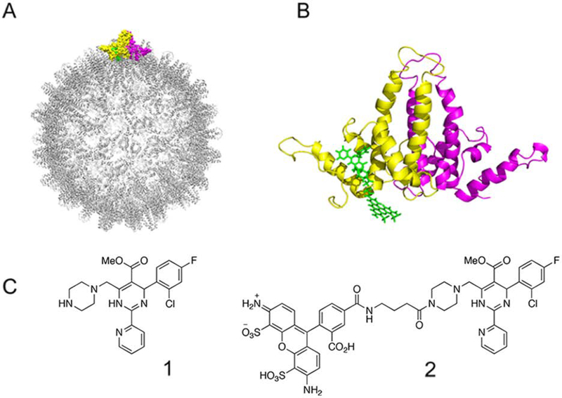
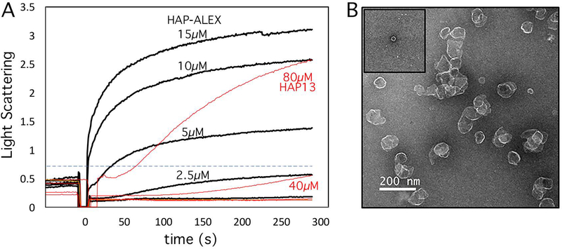
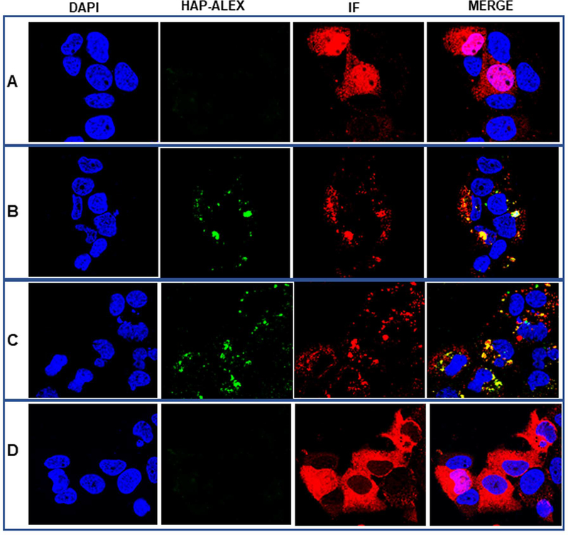
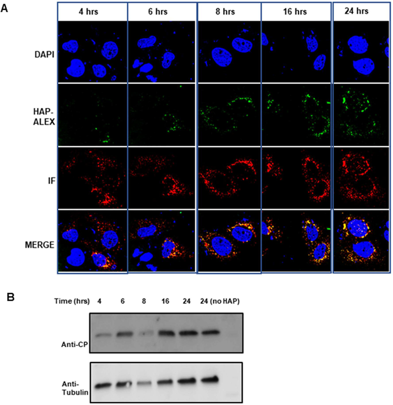
Heteroaryldihydropyrimidines (HAPs) are antiviral small molecules that enhance assembly of HBV core protein (Cp), lead to assembly of empty and defective particles, and suppress viral replication. These core protein allosteric modulators (CpAMs) bind to the pocket at the interface between two Cp dimers and strengthen interdimer interactions. To investigate the CpAM mechanism, we wanted to examine the cellular distributions of Cp and the CpAM itself. For this reason, we developed a fluorescently labeled CpAM, HAP-ALEX. In vitro, HAP-ALEX modulated assembly of purified Cp and at saturating concentrations induced formation of large structures. HAP-ALEX bound capsids and not dimers, making it a capsid-specific molecular tag. HAP-ALEX labeled HBV in transfected cells, with no detectable background with a HAP-insensitive Cp mutant. HAP-ALEX caused redistribution of Cp in a dose-dependent manner consistent with its 0.7 μM EC50, leading to formation of large puncta and an exclusively cytoplasmic distribution. HAP-ALEX colocalized with the redistributed Cp, but large puncta accumulated long before they appeared saturated with the fluorescent CpAM. CpAMs affect HBV assembly and localization; with a fluorescent CpAM both drug and target can be identified.


Figures

Figure 1.
Figure 1.. HAP-ALEX and its putative binding site.
(A) Capsid showing a single, highlighted core protein (Cp) dimer (yellow and pink) with hypothetical bound HAP-ALEX (green). (B) A model of HAP-ALEX in the HAP binding site is consistent with a recent structure with a TAMRA HAP derivative. (C) The parent compound, HAP13 (1), and its ALEXA488 fluorescent derivative, HAP-ALEX (2).
Figure 2.
Figure 2.. Effect of HAP-ALEX on capsid assembly.
(A) HAP-ALEX has a dose dependent effect on the rate and extent of Cp149 assembly as observed by 90° light scattering (in arbitrary units). 10 μM Cp149 dimer was incubated with increasing concentrations of HAP-ALEX (black) or HAP13 (red) for 1 min at 25°C. The unlabeled lines are for 1.25μM HAP-ALEX, 10μM HAP13, and 0 μM HAP13. Assembly was initiated by adding an equal volume of 300 mM NaCl, 50 mM HEPES to attain a final concentration of 5 μM Cp149 dimer, 150 mM NaCl and 50 mM HEPES. The dashed blue line is the scattering of Cp149 capsid formed from 5 μM dimers in 150 mM NaCl; scattering above this intensity is indicative of aberrant assembly or large complexes. Each trace is an average of three independent experiments. (B) Negative stain electron micrograph of assembly products obtained with 15 μM HAP-ALEX; inset shows a normal capsid.
Figure 3.
Figure 3.. Binding of HAP-ALEX to Cp149 capsids.
Titration of 5 μM Cp149 with increasing concentrations of HAP-ALEX was observed by size exclusion chromatography. (A) Absorbance at 280 nm shows that HAP-ALEX increases the amount of capsids and non-capsid polymer at the expense of dimer. (B) Absorbance at 495 nm shows that HAP-ALEX co-elutes with the capsid/non-capsid polymer peak. No detectable HAP-ALEX co-eluted with the dimer peak. HAP-ALEX does adsorb to the column and elutes as a series of peaks at the end of the chromatograph. (C) Analysis of absorbance spectra shows that a maximum of one HAP-ALEX is bound per Cp dimer.
Figure 4.
Figure 4.. Detection of HBV intracellular cores by HAP-ALEX.
HuH7-H1 cells were transfected with an surface protein deficient (HBSAg-) clone of HBV. 3 days post-transfection cells were treated with DMSO or HAP-ALEX for 16 hours following which the cells were fixed and prepared for immunofluorescence (IF). (A) A control wild type transfection with a wild type Cp, treated with DMSO, and stained using a polyclonal anti-Cp (Dako). Note that the HAP-ALEX panel in this row is a blank. (B) A wild type transfection treated with HAP-ALEX and stained using polyclonal anti-Cp. (C) A wild type transfection treated with HAP-ALEX and stained using capsid specific monoclonal Mab3120. (D) Transfection with an HBSAg- HBV clone with the HAP-resistant V124W mutant, treated with HAP-ALEX and stained using polyclonal anti-Cp. As predicted, the mutant failed to bind HAP-ALEX.
Figure 5.
Figure 5.. Detection of HBV intracellular cores by HAP-ALEX:Time course.
HuH7-H1 cells were transfected with the surface protein deficient genomic clone of HBV. 24 hours post-transfection cells were treated with HAP-ALEX for different times (4, 6, 8, 16, 24 hrs) after which the cells were fixed and incubated with anti-Cp (Dako) antibody for immunofluorescence (IF). Samples were visualized using a Leica SP8 confocal microscope. (A) With increased treatment time, HAP-ALEX labelling of Cp puncta increased. (B) Western blot analysis of the amount of intracellular CP during HAP-ALEX treatment for different durations of times (4, 6, 8, 16, 24hrs). HAP-ALEX did not cause any CP degradation during treatment.
Figure 6.
Figure 6.. Detection of HBV intracellular cores by HAP-ALEX: Effect of HAP-ALEX concentration.
(A) HuH7-H1 cells were transfected with the surface protein deficient genomic clone of HBV. 24 hours post-transfection, cells were treated with increasing concentrations of HAP-ALEX (0.1, 0.2, 0.5, 1, 2 μM) for 16 hrs after which the cells were fixed and stained with anti-Cp (Dako) antibody (IF). Samples were visualized using a Leica SP8 confocal microscope. There is no detectable HAP-ALEX signal at concentrations lower than 0.5 μM. At 0.5 μM HAP-ALEX, Cp puncta become noticeably larger and show signs of ALEXA fluorescence. Size of puncta and the HAP-ALEX fluorescence intensity both increase with HAP-ALEX concentration. (B) Western blot analysis of the amount of intracellular CP during various HAP-ALEX treatment.
Figure 7.
Figure 7.. Detection of polymerase defective and empty HBV intracellular cores by HAP-ALEX.
(A) HuH7-H1 cells were transfected with genomic clone of HBV that makes no envelope protein and encodes a Y63F mutant polymerase. These transfections will yield cores that contain pgRNA but are unable to synthesize rcDNA. 3 days post-transfection cells were treated with HAP-ALEX for 16 hours after which the cells were fixed and stained with anti Cp antibodies (Dako) for IF. (B) A parallel control experiment with expression of clone with defective Y63F polymerase and the V124W Cp mutant where Cp does not bind HAP-ALEX. (C) Expression of empty cores was accomplished by transfection of pTruf-HBc which encodes Cp but no other component of HBV. HAP-ALEX binds Cp and induces formation of large puncta under expression conditions that normally yield RNA-filled capsids (A) and empty capsids (C).

Similar articles

Cited by

References

    1. Lavanchy D Hepatitis B virus epidemiology, disease burden, treatment, and current and emerging prevention and control measures. J Viral Hepat 2004, 11, 97. - PubMed
    1. Ott JJ; Stevens GA; Groeger J; Wiersma ST Global epidemiology of hepatitis B virus infection: new estimates of age-specific HBsAg seroprevalence and endemicity. Vaccine 2012, 30, 2212. - PubMed
    1. Lozano R; Naghavi M; Foreman K; Lim S; Shibuya K; Aboyans V; Abraham J; Adair T; Aggarwal R; Ahn SY; et, a. Global and regional mortality from 235 causes of death for 20 age groups in 1990 and 2010: a systematic analysis for the Global Burden of Disease Study 2010. Lancet 2012, 380, 2095. - PMC - PubMed
    1. Buti M HBeAg-positive chronic hepatitis B: why do i treat my patients with Nucleos(t)ide analogs? Liver Int 2014, 34 Suppl 1, 108. - PubMed
    1. Jordheim LP; Durantel D; Zoulim F; Dumontet C Advances in the development of nucleoside and nucleotide analogues for cancer and viral diseases. Nat Rev Drug Discov 2013, 12, 447. - PubMed

Publication types

MeSH terms