. 2019 Jan 2;129(1):252-267.
doi: 10.1172/JCI122064. Epub 2018 Dec 3.

Acetaldehyde dehydrogenase 2 interactions with LDLR and AMPK regulate foam cell formation

Affiliations

Acetaldehyde dehydrogenase 2 interactions with LDLR and AMPK regulate foam cell formation

Shanshan Zhong et al. J Clin Invest. .

Abstract

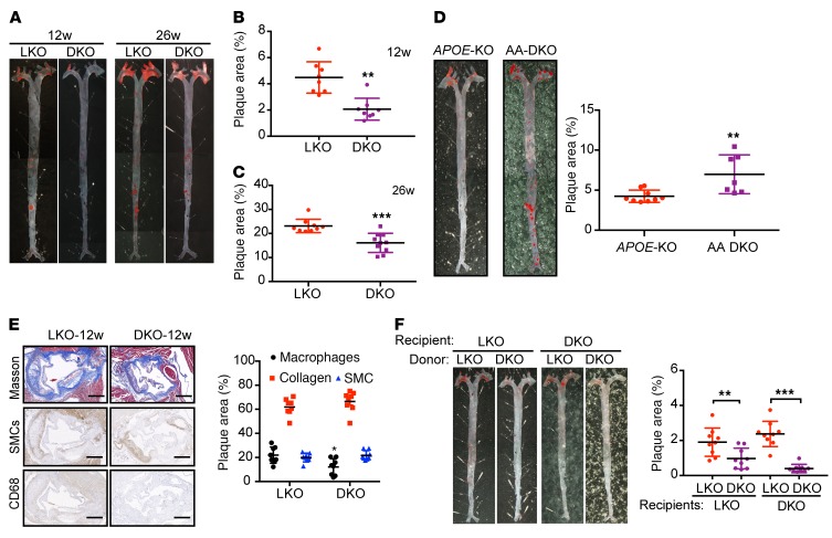
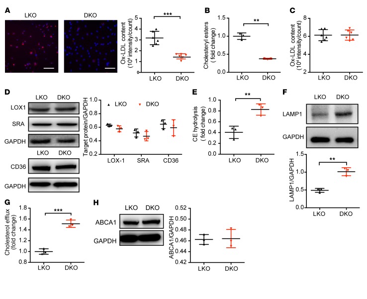
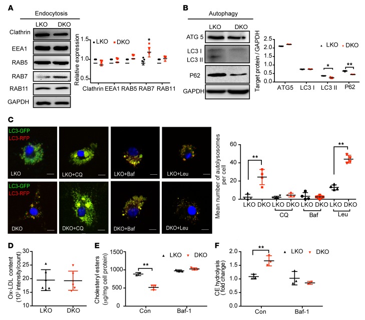
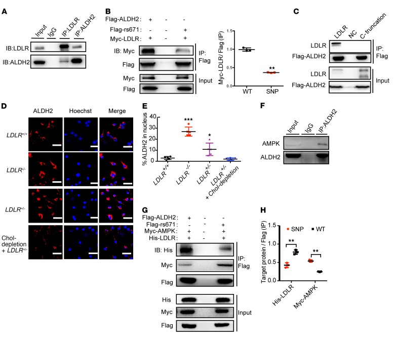
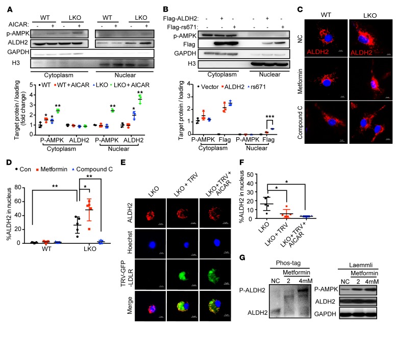
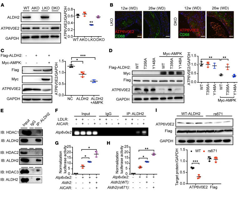
Acetaldehyde dehydrogenase 2 (ALDH2) is a mitochondrial enzyme detoxifying acetaldehyde and endogenous lipid aldehydes; previous studies suggest a protective role of ALDH2 against cardiovascular disease (CVD). Around 40% of East Asians carrying the single nucleotide polymorphism (SNP) ALDH2 rs671 have an increased incidence of CVD. However, the role of ALDH2 in CVD beyond alcohol consumption remains poorly defined. Here we report that ALDH2/LDLR double knockout (DKO) mice have decreased atherosclerosis compared with LDLR-KO mice, whereas ALDH2/APOE-DKO mice have increased atherosclerosis, suggesting an unexpected interaction of ALDH2 with LDLR. Further studies demonstrate that in the absence of LDLR, AMPK phosphorylates ALDH2 at threonine 356 and enables its nuclear translocation. Nuclear ALDH2 interacts with HDAC3 and represses transcription of a lysosomal proton pump protein ATP6V0E2, critical for maintaining lysosomal function, autophagy, and degradation of oxidized low-density lipid protein. Interestingly, an interaction of cytosolic LDLR C-terminus with AMPK blocks ALDH2 phosphorylation and subsequent nuclear translocation, whereas ALDH2 rs671 mutant in human macrophages attenuates this interaction, which releases ALDH2 to the nucleus to suppress ATP6V0E2 expression, resulting in increased foam cells due to impaired lysosomal function. Our studies reveal a novel role of ALDH2 and LDLR in atherosclerosis and provide a molecular mechanism by which ALDH2 rs671 SNP increases CVD.

Keywords: Atherosclerosis; Cardiovascular disease; Cell Biology; Macrophages; Metabolism.


Conflict of interest statement

Conflict of interest: The authors have declared that no conflict of interest exists.

Figures

Figure 1
Figure 1. ALDH2-KO decreases areas of atherosclerotic plaque in LDLR-KO background mice, but increases areas of atherosclerotic plaque in APOE-KO background mice.
(A) Representative en face Sudan IV staining. (B, C) Quantification of plaque areas of aortas from male mice fed WD for 12 weeks (B, n = 8) and 26 weeks (C, n = 9–10, conducted twice). (D) Representative en face Sudan IV staining (n = 7–9) and quantification of Sudan IV–positive areas of aortas from male APOE-KO and APOE/ALDH2-DKO (AA-DKO) mice fed WD for 12 weeks. (E) Representative IHC and quantification of macrophages, collagen, and SMCs after 12 weeks of WD feeding (n = 8). Scale bar: 400 μm. (F) Representative en face Sudan IV staining and quantification of Sudan IV–positive areas of aortas from male LDLR-KO mice (left, n = 9) and LDLR/ALDH2-DKO mice (right, n = 9) transplanted with LDLR-KO and ALDH2/LDLR-DKO bone marrow. Statistical comparisons were made using a 2-tailed Student’s t test. All data are mean ± SD. **P < 0.01, ***P < 0.001. Original magnification for A, D, and F: ×6.3.
Figure 2
Figure 2. ALDH2/LDLR-DKO macrophages exhibit decreased foam cells compared with LDLR-KO macrophages through restoring the lysosomal function and increased CE hydrolysis, without affecting ox-LDL binding and ABCA1 expression.
(A) ALDH2-KO decreases foam cell formation in LDLR-KO (LKO) BMDMs. Quantification is also shown (n = 6). The ox-LDL signals are shown in red and Hoechst signals are shown in blue. Scale bar: 100 μm. (B) CE decreased in ALDH2/LDLR-DKO (DKO) BMDMs compared with LKO macrophages (n = 3). (C) Binding to ox-LDL in LKO and ALDH2/LDLR-DKO BMDMs (n = 6). (D) Expression of LOX1, SRA, and CD36 in LKO and ALDH2/LDLR-DKO BMDMs (n = 3). (E) CE hydrolysis significantly increased in ALDH2/LDLR-DKO BMDMs compared with LKO macrophages (n = 3). (F) Expression of lysosome function marker LAMP1 in LDLR-KO and ALDH2/LDLR-DKO macrophages (n = 3). (G) Cholesterol efflux increased by ALDH2-KO in LDLR-KO BMDMs (n = 3). (H) ABCA1 expression in LKO and DKO BMDMs (n = 3). Statistical comparisons were made using 2-tailed Student’s t test. All data are mean ± SD. **P < 0.01, ***P < 0.001.
Figure 3
Figure 3. LDLR/ALDH2-DKO leads to increased macrophage endocytosis, autophagy, and CE hydrolysis in lysosome compared with LDLR-KO.
(A and B) Endocytosis (A) and autophagy (B) are increased in ALDH2/LDLR-DKO macrophages compared with LDLR-KO (LKO; n = 3). (C) The number of autolysosomes is increased in ALDH2/LDLR-DKO macrophages compared with LKO (n = 4). Scale bars: 5 μm. (DF) Inhibition of autophagy by Baf-A1 treatment diminishes increased foam cell formation (D, n = 6) and cholesteryl ester accumulation (E, n = 3) due to impaired CE hydrolysis (F, n = 3) in LKO macrophages compared with those from DKO. Statistical comparisons were made using 2-tailed Student’s t test. All data are mean ± SD. *P < 0.05, **P < 0.01.
Figure 4
Figure 4. LDLR inhibits but ALDH2 rs671 mutant increases nuclear translocation of ALDH2 through interaction with AMPK.
(A) LDLR directly interacts with ALDH2 in BMDMs (n = 3). (B) ALDH2 rs671 mutant pulls down much less LDLR compared with WT ALDH2 (n = 3). (C) ALDH2 does not bind to LDLR when LDLR C-terminal is truncated (n = 3). (D and E) LDLR gene-dose–dependent inhibition of ALDH2 translocation. (D) LDLR upregulation decreased ALDH2 translocation by cholesterol depletion. Scale bars: 100 μm. Quantification is shown in E (n = 5). (F) ALDH2 directly binds to AMPK in LDLR-KO BMDMs. (G and H) ALDH2 rs671 mutant pulls down more AMPK compared with WT ALDH2 by cotransfection of Flag-tagged ALDH2, Myc-tagged AMPK, and His-tagged LDLR (G) and quantification (H, n = 3). Statistical comparisons were made using 2-tailed Student’s t test (B and H) or ANOVA (E). All data are mean ± SD. *P < 0.05, **P < 0.01, ***P < 0.001.
Figure 5
Figure 5. AMPK phosphorylates ALDH2 and promotes ALDH2 translocation in the absence of LDLR or ALDH2 rs671 mutant.
(A) AMPK activation promotes ALDH2 nuclear translocation in LDLR-KO BMDMs by cellular fractionation (LKO; n = 3). (B) ALDH2 rs671 mutant increases the translocation of ALDH2 in 293T cells (n = 3). (C) AMPK activation leads to ALDH2 nuclear translocation, whereas inhibition of AMPK blocks nuclear translocation of ALDH2. Scale bars: 5 μm. (D) Quantification of ALDH2 in the nucleus (n = 5). (E) LDLR blocks the translocation of ALDH2 even AMPK activation. Scale bars: 5 μm. Quantification shown in F (n = 5). (G) AMPK activation leads to a dose-dependent increase of ALDH2 phosphorylation in the absence of LDLR in LKO macrophages by SuperSep Phos-tag SDS-PAGE. Statistical comparisons were made using ANOVA. All data are mean ± SD. *P < 0.05, **P < 0.01, ***P < 0.001.
Figure 6
Figure 6. AMPK regulates lysosomal function, endocytosis, autophagy, and foam cell formation in an LDLR-dependent manner.
(A) LDLR-KO increases the AMPK activation in BMDMs (n = 3). (B) Ratio of AMP/ATP in macrophages from LDLR-KO and WT treated with ox-LDL (n = 3). (C) LDLR inhibits the binding of ALDH2 and AMPK in macrophages. (DF) AMPK and ALDH2 prefer to bind to LDLR. LDLR pulls down AMPK and ALDH2 (D). AMPK pulls down LDLR but not ALDH2 (E). ALDH2 pulls down LDLR but not AMPK (F). (G) AMPK activation by metformin decreases LAMP1 expression, whereas AMPK inhibition by compound C leads to increased LAMP1 expression in LDLR-KO BMDMs (LKO; n = 3). (H and I) AMPK activation by metformin decreases endocytosis (H) and autophagy (I), whereas AMPK inhibition by compound C leads to increased endocytosis (H) and autophagy (I) in LKO BMDMs (n = 3). (J) AMPK activation leads to increased foam cell formation, whereas AMPK inhibition results in decreased foam cell formation (n = 5). (K) AMPK activation decreases CE hydrolysis, whereas AMPK inhibition increases CE hydrolysis (n = 3). Statistical comparisons were made using 2-tailed Student’s t test (A and B) or ANOVA (G, HK). All data are mean ± SD. *P < 0.05, **P < 0.01, ***P < 0.001.
Figure 7
Figure 7. Nuclear translocated ALDH2 regulates the transcription of ATP6V0E2, a critical protein for lysosomal function, endocytosis, and autophagy, and ALDH2 rs671 polymorphism decreases ATP6V0E2 expression.
(A) ALDH2/LDLR-DKO leads to significant upregulation of ATP6V0E2 expression compared with LDLR-KO (LKO; n = 3). (B) ATP6V0E2 (red) colocalizes with macrophages (CD68, green) and ATP6V0E2 expression is significantly increased in the aorta of DKO mice. Scale bars: 100 μm. (C) Overexpressed AMPK and ALDH2 decrease ATP6V0E2 expression in 293T cells (n = 3). (D) ALDH2 T356A not Y148A mutant rescues decreased ATP6V0E2 expression, which is caused by overexpressed ALDH2 and AMPK (n = 3). (E) Nuclear translocated ALDH2 binds to HDAC3. (F and G) Nuclear translocated ALDH2 regulates transcription of ATP6V0E2. In the absence of LDLR, nuclear translocated ALDH2 binds to ATP6V0E2 promoter (F) and regulates transcriptional activity (G), which is enhanced by AMPK activation (n = 3). (H) ALDH2 rs671 enhanced transcriptional activity of ATP6V0E2. (I) The rs671 mutant decreased ATP6V0E2 protein expression (n = 3). Statistical comparisons were made using 2-tailed Student’s t test (I) or ANOVA (A, C, D, G, and H). All data are mean ± SD. *P < 0.05, **P < 0.01, ***P < 0.001.
Figure 8
Figure 8. Human macrophages from subjects carrying ALDH2 rs671 SNP have increased foam cell formation caused by the downregulation of ATP6V0E2, and autophagy due to the increased nuclear translocation of ALDH2.
(A) Macrophages from ALDH2*2/2*1 (n = 10) have lower ATP6V0E2 mRNA levels than those from ALDH2*1 (n = 16). (B) ALDH2 rs671 mutant increased autophagy in human macrophages treated with ox-LDL (n = 3). (C) ALDH2 rs671 mutant decreases CE hydrolysis in human macrophages (ALDH2*1, n = 16; ALDH2*2/2*1, n = 10). (D) ALDH2 rs671 SNP increases ALDH2 translocation in human macrophages treated with metformin (n = 5). Scale bars: 5 μm. Statistical comparisons were made using 2-tailed Student’s t test. All data are mean ± SD. *P < 0.05, **P < 0.01, ***P < 0.001.
Figure 9
Figure 9. ALDH2 regulates macrophage foam cell formation through interacting with LDLR and AMPK.
The translocation of AMPK phosphorylated ALDH2 to the nucleus to regulate expression of ATP6V0E2, a critical protein for lysosomal function, endocytosis, and autophagy.

Comment in

References

    1. Weber C, Noels H. Atherosclerosis: current pathogenesis and therapeutic options. Nat Med. 2011;17(11):1410–1422. doi: 10.1038/nm.2538. - DOI - PubMed
    1. Moore KJ, Tabas I. Macrophages in the pathogenesis of atherosclerosis. Cell. 2011;145(3):341–355. doi: 10.1016/j.cell.2011.04.005. - DOI - PMC - PubMed
    1. Zhao B, Song J, Chow WN, St Clair RW, Rudel LL, Ghosh S. Macrophage-specific transgenic expression of cholesteryl ester hydrolase significantly reduces atherosclerosis and lesion necrosis in Ldlr mice. J Clin Invest. 2007;117(10):2983–2992. doi: 10.1172/JCI30485. - DOI - PMC - PubMed
    1. Ouimet M, et al. microRNA-33 regulates macrophage autophagy in atherosclerosis. Arterioscler Thromb Vasc Biol. 2017;37(6):1058–1067. doi: 10.1161/ATVBAHA.116.308916. - DOI - PMC - PubMed
    1. Sergin I, Evans TD, Razani B. Degradation and beyond: the macrophage lysosome as a nexus for nutrient sensing and processing in atherosclerosis. Curr Opin Lipidol. 2015;26(5):394–404. doi: 10.1097/MOL.0000000000000213. - DOI - PMC - PubMed

Publication types

MeSH terms

Substances