. 2018 Oct 30;37(1):263.
doi: 10.1186/s13046-018-0919-8.

ATF3 inhibits the tumorigenesis and progression of hepatocellular carcinoma cells via upregulation of CYR61 expression

Affiliations

ATF3 inhibits the tumorigenesis and progression of hepatocellular carcinoma cells via upregulation of CYR61 expression

Cong Chen et al. J Exp Clin Cancer Res. .

Abstract

Background: Hepatocellular carcinoma (HCC) is one of the most common malignant cancers with a high incidence and high mortality in East Asia. Identifying biomarkers and clarifying the regulatory mechanisms of HCC are of great importance. Herein, we report the role and mechanism of activating transcription factor 3 (ATF3), a member of the ATF/cAMP-responsive element-binding protein family of transcription factors in HCC.

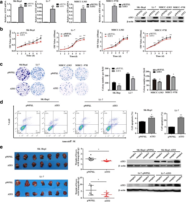
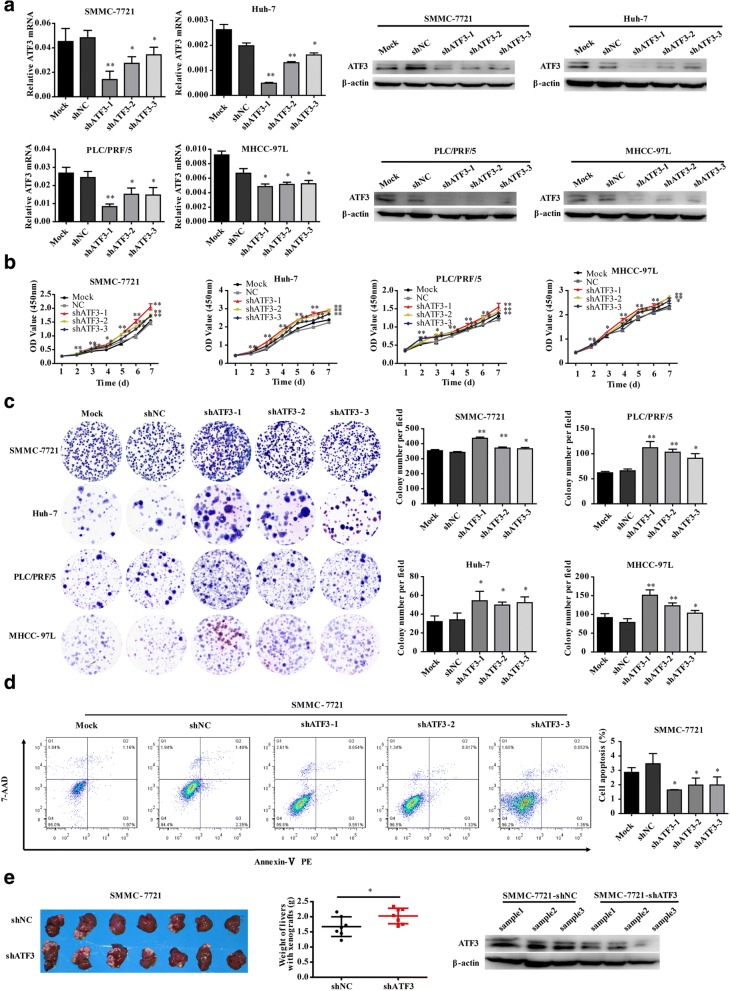
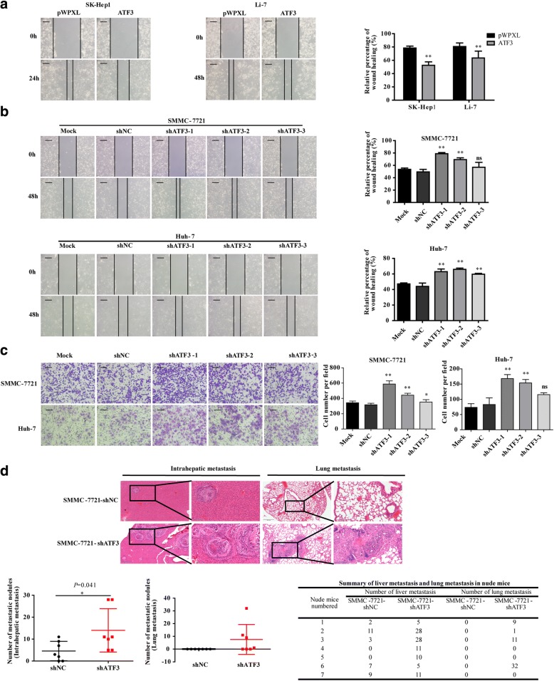
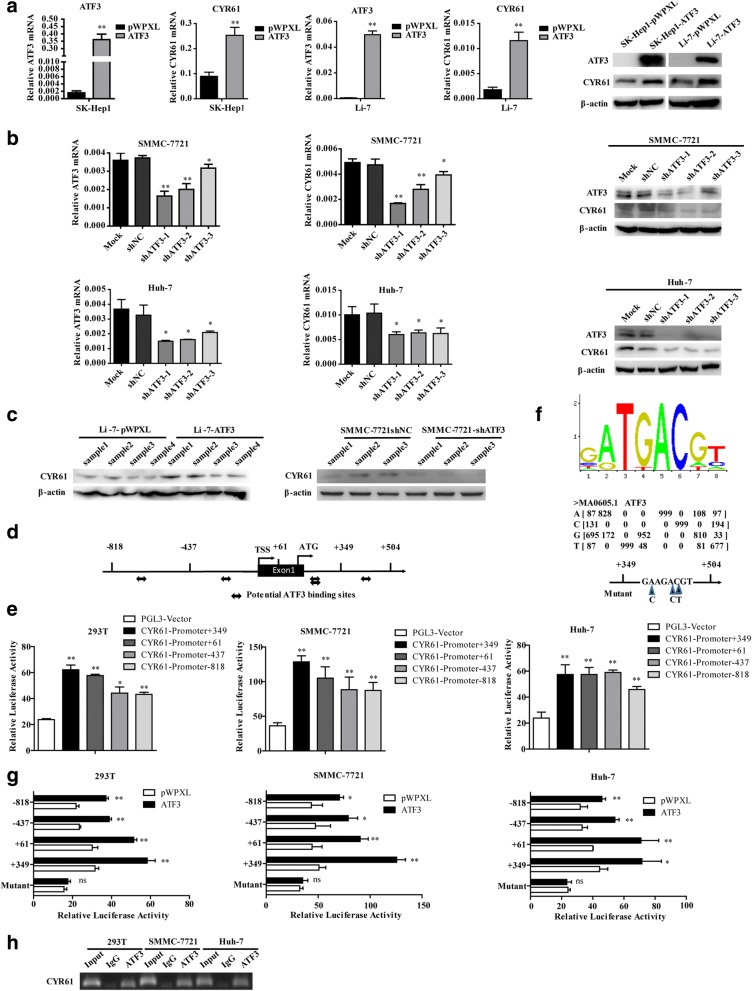
Methods: ATF3 overexpression vector and shRNAs were transfected into HCC cancer cells to upregulate or downregulate ATF3 expression. In vitro and in vivo assays were performed to investigate the functional role of ATF3 in hepatocellular carcinoma. RNA-Seq was performed to screen the differentially expressed genes downstream of ATF3. The dual-luciferase reporter assay, chromatin immunoprecipitation (Ch-IP) analysis and functional rescue experiments were used to confirm the target gene regulated by ATF3. Tissue microarrays (TMAs) comprising 236 human primary HCC tissues were obtained and immunohistochemical staining were carried out to analyze the clinical significance of ATF3.

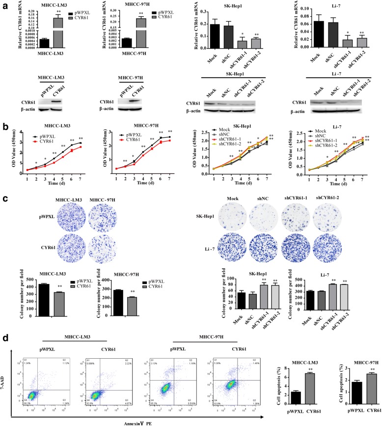
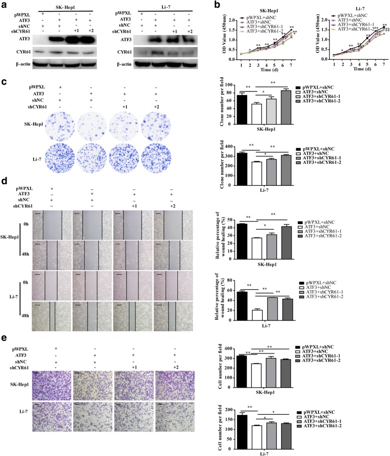
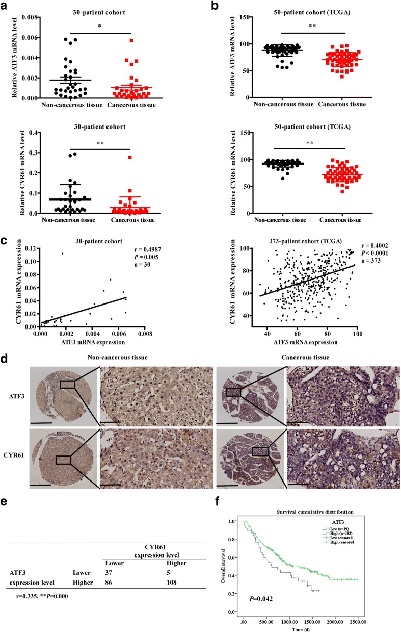
Results: The results indicate that ATF3 significantly inhibited the proliferation and mobility of HCC cells both in vitro and in vivo. Cysteine-rich angiogenic inducer 61 (CYR61) is a key target for transcriptional regulation by ATF3. Both ATF3 and CYR61 were consistently downregulated in human HCC tissues, and their expression levels were significantly and positively correlated with each other.

Conclusions: Our findings indicate that ATF3 functions as a tumor suppressor in HCC through targeting and regulating CYR61.

Keywords: ATF3; CYR61; Hepatocellular carcinoma.


Conflict of interest statement

Ethics approval and consent to participate

All animal procedures were performed under the guidelines of the Shanghai Medical Experimental Animal Care Commission. The study was approved by the Chinese Ethical Review Committee and signed informed consent was obtained from each patient.

Consent for publication

All the patients that involved in the study have given their consent to publish their individual data.

Competing interests

The authors declare that they have no competing interests.

Publisher’s Note

Springer Nature remains neutral with regard to jurisdictional claims in published maps and institutional affiliations.

Figures

Fig. 1
Fig. 1
ATF3 inhibited HCC cell proliferation in vitro and tumorigenesis in vivo. a As a quality control, in vitro ATF3 mRNA and protein expression was detected by qRT-PCR and western blot, respectively, in SK-Hep1, Li-7, MHCC-LM3 and MHCC-97H cells with ectopic ATF3 expression via the lentiviral vector (pWPXL). b The cell proliferative ability of HCC cells with ATF3 overexpression was inhibited as indicated by the CCK-8 assay. c The colony forming ability of HCC cells with ATF3 overexpression was reduced. d The percent of cell apoptosis with ATF3 overexpression in SK-Hep1 and Li-7 was increased. e Images of liver tissues collected from nude mice with tumor xenografts derived from SK-Hep1 (left top panel) and Li-7 (left bottom panel) cells with stable overexpression of ATF3. The livers with xenografts were weighed and compared (middle top and bottom panel). ATF3 protein expression in tissue samples of the xenografts was detected by western blot (right top and bottom panel). β-Actin was used as a loading control. The bar graphs in (a), (b), (c) and (d) represent the quantitative data from three independent experiments. Unpaired Student’s t-test was used for statistical analysis, and the data are shown as the mean ± S.D. *P < 0.05 and **P < 0.01
Fig. 2
Fig. 2
Silencing ATF3 promoted tumor cell proliferation in vitro and in vivo. a As a quality control, in vitro ATF3 mRNA and protein expression was detected by qRT-PCR and western blot, respectively, in SMMC-7721, Huh-7, PLC/PRF/5 and MHCC-97 L cells with stable knockdown of ATF3. Mock and shNC-expressing cells served as blank and negative control groups, respectively. Compared to the NC and mock groups, SMMC-7721, Huh-7, PLC/PRF/5 and MHCC-97 L cells expressing shATF3 showed increased cell proliferation (b) and colony forming ability (c). d The percent of cell apoptosis with ATF3 knockdown in SMMC-7721 was decreased. e Images of liver tissues collected from nude mice with tumor xenografts derived from SMMC-7721 (left panel) cells with stable knockdown of ATF3. The livers with xenografts were weighed (middle panel). The ATF3 protein level in tissue samples from the xenografts was detected by western blot (right panel). β-Actin was used as a loading control. The bar graphs in (a), (b), (c) and (d) represent the quantitative data from three independent experiments. Unpaired Student’s t-test was used for statistical analysis, and the data are shown as the mean ± S.D. *P < 0.05 and **P < 0.01
Fig. 3
Fig. 3
ATF3 suppressed HCC cell mobility in vitro and metastasis in vivo. a Overexpression of ATF3 suppressed HCC cell migration in vitro as assessed by the wound healing assay (scale bar, 200 μm). b Knockdown of ATF3 (shATF3–1, − 2, − 3) promoted HCC cell migration in vitro as assessed by the wound healing assay (scale bar, 200 μm). c Silencing ATF3 promoted HCC cell invasion in vitro (scale bar, 100 μm). The bar graphs in (a), (b) and (c) represent quantitative data from three replicates. d Representative images show the intrahepatic metastatic and lung metastatic nodules derived from SMMC-7721 cells with silenced ATF3 and those derived from the control cells (left images, scale bar, 500 μm; right images, scale bar, 100 μm). The numbers of intrahepatic metastatic and lung metastatic nodules are presented in the bottom panel (n = 7). Unpaired Student’s t-test was used for statistical analysis, and the data are shown as the mean ± S.D. *P < 0.05, **P < 0.01 and ns: no significance
Fig. 4
Fig. 4
ATF3 upregulated CYR61 by directly binding to its consensus binding sequence on the CYR61 gene. a qRT-PCR and western blot showed that SK-Hep1 and Li-7 cells overexpressing ATF3 had increased CYR61 mRNA and protein expression. b qRT-PCR and western blot showed that SMMC-7721 and Huh-7 cells with ATF3 knockdown had decreased CYR61 mRNA and protein expression. c Upregulated expression of CYR61 protein in tissue samples from xenografts was detected by western blot. d Potential ATF3 binding sites next to the transcriptional start site of the CYR61 sequence were identified with the JASPAR database (http://jaspar.genereg.net/). e The relative luciferase activities of the full CYR61 sequence and the truncated construct in 293 T, SMMC-7721 and Huh-7 cells. f The sequence logo of a potential ATF3 binding site in JASPAR and a diagram of mutant sites in the CYR61 sequence. g The relative luciferase activities of the truncated and mutant constructs of the CYR61 sequence in 293 T, SMMC-7721 and Huh-7 cells transfected with either ATF3 or pWPXL. h Assessment of ATF3 binding to the CYR61 sequence in 293 T, SMMC-7721 and Huh-7 cells was performed by Ch-IP using an antibody against ATF3 and a negative control (IgG). Agarose gel electrophoresis was used to analyze the crosslinking status. Data are shown as the mean ± S.D. from experiments with three replicates. *P < 0.05, **P < 0.01 and ns: no significance
Fig. 5
Fig. 5
CYR61 suppressed HCC cell proliferation in vitro. a As a quality control, in vitro CYR61 mRNA and protein expression was detected by qRT-PCR and western blot, respectively, in MHCC-LM3 and MHCC-97H cells with ectopic CYR61 expression via the lentiviral vector (pWPXL) and SK-Hep1, Li-7 cells with silencing of CYR61. The cell proliferative ability of HCC cells with CYR61 overexpression and knockdown were examined by the CCK-8 assay (b) and the colony formation assay (c). d The percent of cell apoptosis with CYR61 overexpression was increased. The bar graphs in (a), (b), (c) and (d) represent the quantitative data from three independent experiments. Unpaired Student’s t-test was used for statistical analysis, and the data are shown as the mean ± S.D. *P < 0.05 and **P < 0.01
Fig. 6
Fig. 6
Knockdown CYR61 in HCC cells overexpressing ATF3 rescued the suppressive effects of ATF3 in vitro. a Western blot of CYR61 protein expression in ATF3-overexpressing HCC cells with CYR61 knockdown (shCYR61–1, − 2). β-Actin was used as a loading control. The influence of CYR61 knockdown on the inhibitory effects of ATF3 on cell growth (b), colony formation (d), migration (d) and invasion (e) in vitro were detected by the CCK-8 assay, colony formation assay, wound healing assay (scale bar, 200 μm) and transwell invasion assay (scale bar, 100 μm), respectively. The bar graphs in (b), (c), (d) and (e) represent quantitative data from three replicates. Data are shown as the mean ± S.D. from experiments with three replicates. *P < 0.05 and **P < 0.01
Fig. 7
Fig. 7
ATF3 expression was positively correlated with CYR61 expression in primary HCC tissues. The mRNA expression of ATF3 and CYR61 in 30 pairs of HCC and noncancerous liver tissues was detected by qRT-PCR (a), which was also used to analyze 50 pairs of cancerous and noncancerous liver tissues from the TCGA (b). c The correlation between ATF3 and CYR61 mRNA levels in 30 paired cancerous/noncancerous liver tissues from primary HCC patients and 373 paired samples from the TCGA cohort. The Pearson correlation coefficients (r) and p value are indicated. d Representative images of ATF3 and CYR61 protein staining by IHC in cancerous /noncancerous liver tissues (scale bar, 100 μm.). e The positive correlation between ATF3 and CYR61 protein levels in 236 human primary HCC tissues is presented. r, Pearson correlation coefficient; p, p value. f Kaplan–Meier analysis of overall survival in 222 HCC patients, as stratified by ATF3 expression. The log-rank test was used to determine statistical significance. *P < 0.05 and **P < 0.01

References

    1. Siegel RL, Miller KD, Jemal A. Cancer statistics, 2018. CA Cancer J Clin. 2018;68(1):7–30. doi: 10.3322/caac.21442. - DOI - PubMed
    1. Chen W, Zheng R, Baade PD, Zhang S, Zeng H, Bray F, Jemal A, Yu XQ, He J. Cancer statistics in China, 2015. CA Cancer J Clin. 2016;66(2):115–132. doi: 10.3322/caac.21338. - DOI - PubMed
    1. Liu Y, Gao F, Jiang H, Niu L, Bi Y, Young CY, Yuan H, Lou H. Induction of DNA damage and ATF3 by retigeric acid B, a novel topoisomerase II inhibitor, promotes apoptosis in prostate cancer cells. Cancer Lett. 2013;337(1):66–76. doi: 10.1016/j.canlet.2013.05.022. - DOI - PubMed
    1. Kaneko A, Kiryu-Seo S, Matsumoto S, Kiyama H. Damage-induced neuronal endopeptidase (DINE) enhances axonal regeneration potential of retinal ganglion cells after optic nerve injury. Cell Death Dis. 2017;8(6):e2847. doi: 10.1038/cddis.2017.212. - DOI - PMC - PubMed
    1. Wang Z, Kim J, Teng Y, Ding HF, Zhang J, Hai T, Cowell JK, Yan C. Loss of ATF3 promotes hormone-induced prostate carcinogenesis and the emergence of CK5(+)CK8(+) epithelial cells. Oncogene. 2016;35(27):3555–3564. doi: 10.1038/onc.2015.417. - DOI - PMC - PubMed

MeSH terms

Substances