. 2018 Oct 31;9(1):292.
doi: 10.1186/s13287-018-1027-6.

Identifying deer antler uhrf1 proliferation and s100a10 mineralization genes using comparative RNA-seq

Affiliations

Identifying deer antler uhrf1 proliferation and s100a10 mineralization genes using comparative RNA-seq

Dai Fei Elmer Ker et al. Stem Cell Res Ther. .

Abstract

Background: Deer antlers are bony structures that re-grow at very high rates, making them an attractive model for studying rapid bone regeneration.

Methods: To identify the genes that are involved in this fast pace of bone growth, an in vitro RNA-seq model that paralleled the sharp differences in bone growth between deer antlers and humans was established. Subsequently, RNA-seq (> 60 million reads per library) was used to compare transcriptomic profiles. Uniquely expressed deer antler proliferation as well as mineralization genes were identified via a combination of differential gene expression and subtraction analysis. Thereafter, the physiological relevance as well as contributions of these identified genes were determined by immunofluorescence, gene overexpression, and gene knockdown studies.

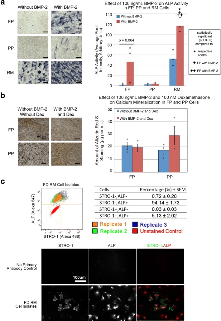
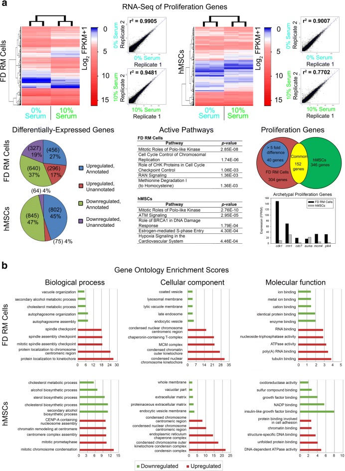
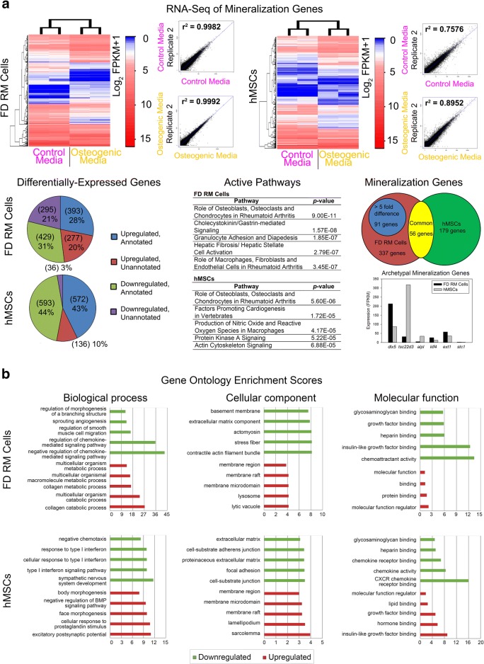
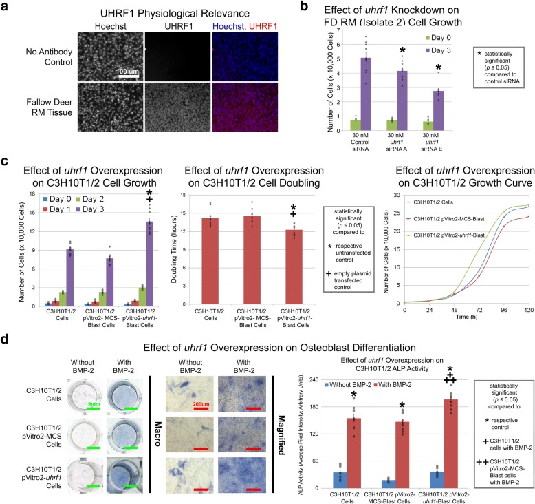
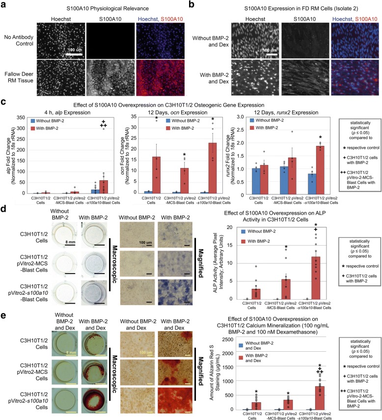
Results: Cell characterization studies showed that in vitro-cultured deer antler-derived reserve mesenchyme (RM) cells exhibited high osteogenic capabilities and cell surface markers similar to in vivo counterparts. Under identical culture conditions, deer antler RM cells proliferated faster (8.6-11.7-fold increase in cell numbers) and exhibited increased osteogenic differentiation (17.4-fold increase in calcium mineralization) compared to human mesenchymal stem cells (hMSCs), paralleling in vivo conditions. Comparative RNA-seq identified 40 and 91 previously unknown and uniquely expressed fallow deer (FD) proliferation and mineralization genes, respectively, including uhrf1 and s100a10. Immunofluorescence studies showed that uhrf1 and s100a10 were expressed in regenerating deer antlers while gene overexpression and gene knockdown studies demonstrated the proliferation contributions of uhrf1 and mineralization capabilities of s100a10.

Conclusion: Using a simple, in vitro comparative RNA-seq approach, novel genes pertinent to fast bony antler regeneration were identified and their proliferative/osteogenic function was verified via gene overexpression, knockdown, and immunostaining. This combinatorial approach may be applicable to discover unique gene contributions between any two organisms for a given phenomenon-of-interest.

Keywords: Bone regeneration; Comparative RNA-seq; Deer antler; s100a10; uhrf1.


Conflict of interest statement

Ethics approval

Ethics approval for the harvesting of deer antlers was obtained from Stanford University’s Administrative Panel on Laboratory Animal Care (APLAC 28057).

Consent for publication

Not applicable.

Competing interests

The authors declare that they have no competing interests.

Publisher’s Note

Springer Nature remains neutral with regard to jurisdictional claims in published maps and institutional affiliations.

Figures

Fig. 1
Fig. 1
Characterization of in vitro-cultured FD-derived cells. a FP and RM cells (isolate 2) cultured with 100 ng/mL BMP-2 for 6 days exhibited increased ALP activity relative to their respective control whereas PP cells (isolate 2) did not. Semi-quantification of ALP activity in FP, PP, and RM cells. b FP and PP cells (isolate 2) cultured with 100 ng/mL BMP-2 and 100 nM dexamethasone for 24 days did not exhibit increased Alizarin Red S staining relative to their respective control. Quantification of Alizarin Red S staining in FP and PP cells. c FACS analysis of RM cells. The percentage of cells that were negative for STRO1 and ALP, negative for STRO1 but positive for ALP, negative for ALP but positive for STRO1, and positive for both STRO1 and ALP were 0.28–1.25%, 91.83–97.53%, 0.004–0.10%, and 1.20–7.89%, respectively. STRO1 and ALP immunofluorescence staining in RM cells. Green, STRO1-positive cells. Red, ALP-positive cells. Scale bars as indicated. Data were from n = 3 isolates (three independent experiments with nine replicates per isolate for ALP and mineralization studies and one independent experiment with three replicates per isolate for FACs studies). Gray circles indicate observed data points. Error bars indicate standard error of mean or SEM. Statistical significance as indicated
Fig. 2
Fig. 2
RM cells exhibit increased proliferation and osteogenic differentiation compared to hMSCs. a RM cells exhibited increased proliferation relative to hMSCs. b Cell cycle analysis showed an increased proportion of RM cells undergoing cell division relative to hMSCs. c RM cells were capable of chondrogenic but not adipogenic differentiation. d RM cells (isolate 2) cultured with 100 ng/mL BMP-2 for 6 days exhibited increased osteogenic gene expression relative to their respective control. e RM cells cultured with 100 ng/mL BMP-2 and 100 nM dexamethasone for 24 days exhibited increased Alizarin Red S staining relative to hMSCs. Scale bars as indicated. Data were from n = 3 isolates (three independent experiments with nine replicates per isolate for proliferation and chondrogenic, adipogenic, and mineralization studies and one independent experiment with three replicates per isolate for cell cycle studies) or n = 1 isolate (two independent experiments with six replicates for osteogenic gene expression studies). Gray circles indicate observed data points. Error bars indicate SEM. Statistical significance as indicated
Fig. 3
Fig. 3
RNA-seq analysis of RM cells and hMSCs under proliferation and mineralization conditions. a RNA-seq analysis of RM cells (isolate 2) and hMSCs (isolate 24268) under serum-free (0% serum) and serum-containing (10% serum) conditions identified 40 candidate proliferation genes. Scatterplots indicate the correlation (r2) between replicates for each condition. FPKM, fragments per kilobase of transcript per million mapped reads. b Gene ontology enrichment analysis of RM cells (isolate 2) and hMSCs (isolate 24268) under proliferation conditions. Graphs indicate the top 5 upregulated (red) and downregulated (green) biological processes, cellular components, and molecular functions. Data were from n = 1 isolate (one independent experiment with two replicates per group)
Fig. 4
Fig. 4
RNA-seq analysis of RM cells and hMSCs under proliferation and mineralization conditions. a RNA-seq analysis of RM cells (isolate 2) and hMSCs (isolate 24268) under control (0 ng/mL BMP-2 and 0 nM dexamethasone) and osteogenic (100 ng/mL BMP-2 and 100 nM dexamethasone) conditions identified 91 candidate mineralization genes. Scatterplots indicate the correlation (r2) between replicates for each condition. FPKM, fragments per kilobase of transcript per million mapped reads. b Gene ontology enrichment analysis of RM cells (isolate 2) and hMSCs (isolate 24268) under mineralization conditions. Graphs indicate the top 5 upregulated (red) and downregulated (green) biological processes, cellular components, and molecular functions. Data were from n = 1 isolate (one independent experiment with two replicates per group)
Fig. 5
Fig. 5
Identification of uhrf1 as a uniquely expressed proliferation gene using in vitro comparative RNA-seq. a UHRF1 immunofluorescence staining in regenerating deer antler tissue. b RM cells cultured with 30 nM uhrf1 siRNAs for 3 days exhibited decreased proliferation relative to mock-transfected control. c C3H10T1/2 cells stably transfected with uhrf1 exhibited increased proliferation relative to untransfected control and empty plasmid control. C3H10T1/2 cells stably transfected with uhrf1 maintained contact inhibition. Representative growth curves are shown. d C3H10T1/2 cells stably transfected with uhrf1 and cultured with 100 ng/mL BMP-2 for 6 days exhibited increased ALP activity relative to untransfected control and empty plasmid control. Scale bars as indicated. Data were from n = 3 isolates (an independent herd for antler immunofluorescence studies) or n = 3 independent experiments with nine replicates per group for uhrf1 knockdown and overexpression proliferation and osteoblast differentiation studies. Gray circles indicate observed data points. Error bars indicate SEM. Statistical significance as indicated
Fig. 6
Fig. 6
Identification of s100a10 as a uniquely expressed mineralization gene using in vitro comparative RNA-seq. a S100A10 immunofluorescence staining in regenerating deer antler tissue. b RM cells (isolate 2) cultured with 100 ng/mL BMP-2 and 100 nM dexamethasone exhibited increased S100A10 expression relative to control. c C3H10T1/2 cells stably transfected with s100a10 and cultured with 100 ng/mL BMP-2 for 4 h exhibited increased alp gene expression relative to untransfected control and empty plasmid control. C3H10T1/2 cells stably transfected with s100a10 and cultured with 100 ng/mL BMP-2 for 12 days exhibited increased ocn and runx2 gene expression relative to their respective control. d C3H10T1/2 cells stably transfected with s100a10 and cultured with 100 ng/mL BMP-2 for 4 days exhibited increased ALP activity relative to untransfected control. e C3H10T1/2 cells stably transfected with s100a10 and cultured in the presence of 100 ng/mL BMP-2 and 100 nM dexamethasone exhibited increased Alizarin Red S staining relative to untransfected control and empty plasmid control. Scale bars as indicated. Data were from n = 3 isolates (an independent herd for antler immunofluorescence studies), n = 2–3 independent experiments with 4–10 replicates per group for osteogenic gene expression studies, n = 3 independent experiments with 9 replicates per group for s100a10 overexpression ALP studies, and n = 5 independent experiments with 15 replicates per group for s100a10 overexpression mineralization studies. Gray circles indicate observed data points. Error bars indicate SEM. Statistical significance as indicated

References

    1. Gómez JA, Ceacero F, Landete-Castillejos T, Gaspar-López E, García AJ, Gallego L. Factors affecting antler investment in Iberian red deer. Anim Prod Sci. 2012;52:867–873. doi: 10.1071/AN11316. - DOI
    1. Huxley JS. The relative size of antlers in deer. Proc Zool Soc London. 1931;101:819–864. doi: 10.1111/j.1096-3642.1931.tb01047.x. - DOI
    1. Goss RJ. Deer antlers. Regeneration, function and evolution. New York: Academic Press; 1983.
    1. Kierdorf U, Kierdorf H. Deer antlers - a model of mammalian appendage regeneration: an extensive review. Gerontology. 2011;57:53–65. doi: 10.1159/000300565. - DOI - PubMed
    1. Li C. Histogenetic aspects of deer antler development. Front Biosci. 2013;5:479–489. doi: 10.2741/E629. - DOI - PubMed

Publication types

Substances