. 2018 Oct 30;11(554):eaar3924.
doi: 10.1126/scisignal.aar3924.

RSK2 contributes to myogenic vasoconstriction of resistance arteries by activating smooth muscle myosin and the Na+/H+ exchanger

Affiliations

RSK2 contributes to myogenic vasoconstriction of resistance arteries by activating smooth muscle myosin and the Na+/H+ exchanger

Mykhaylo V Artamonov et al. Sci Signal. .

Abstract

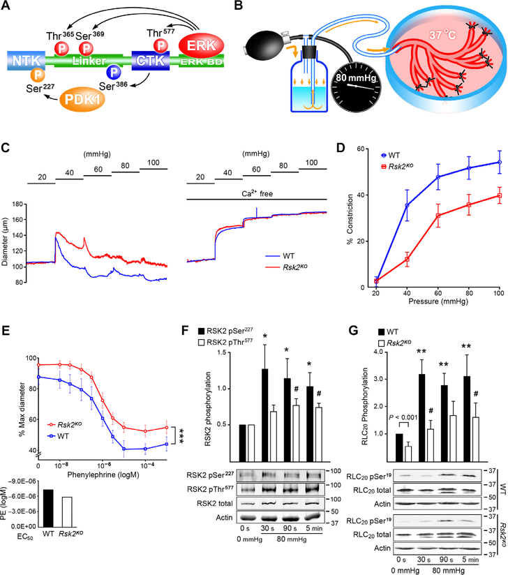
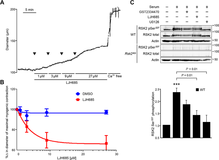
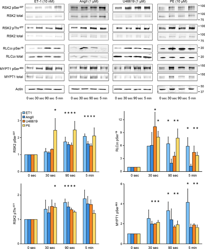
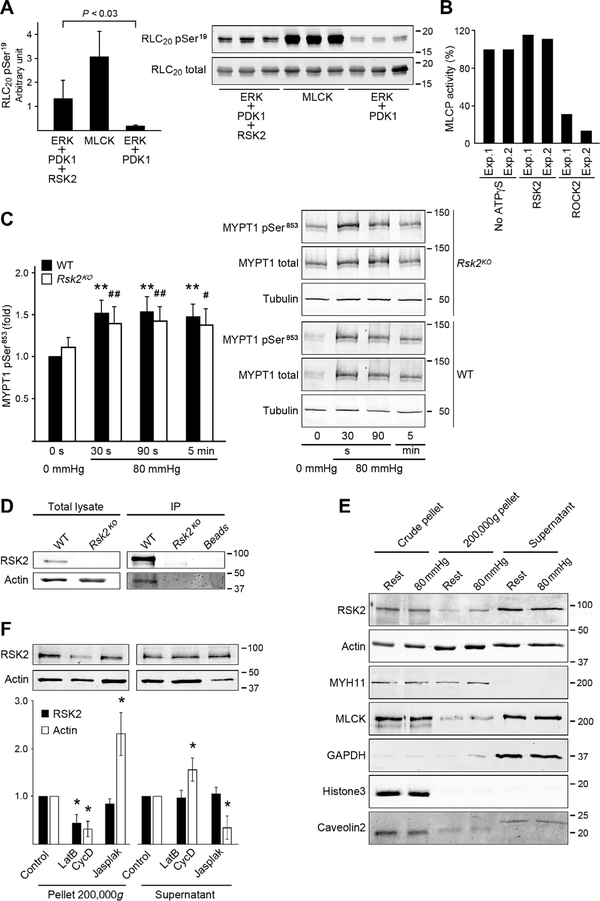
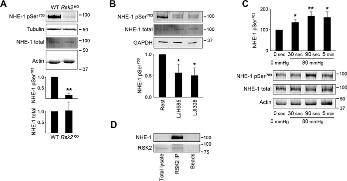
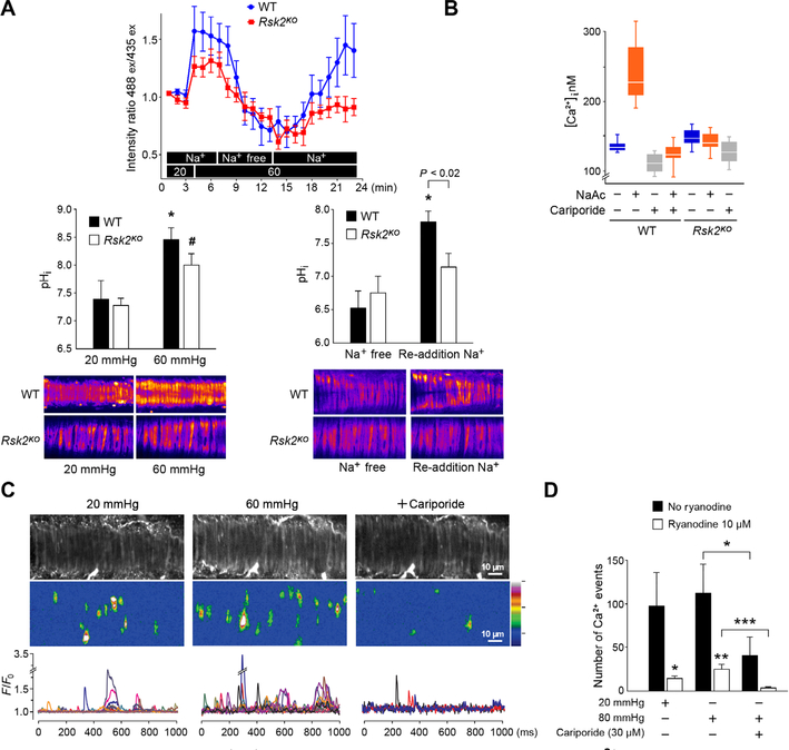
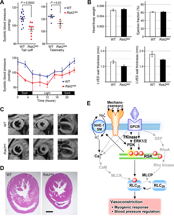
Smooth muscle contraction is triggered when Ca2+/calmodulin-dependent myosin light chain kinase (MLCK) phosphorylates the regulatory light chain of myosin (RLC20). However, blood vessels from Mlck-deficient mouse embryos retain the ability to contract, suggesting the existence of additional regulatory mechanisms. We showed that the p90 ribosomal S6 kinase 2 (RSK2) also phosphorylated RLC20 to promote smooth muscle contractility. Active, phosphorylated RSK2 was present in mouse resistance arteries under normal basal tone, and phosphorylation of RSK2 increased with myogenic vasoconstriction or agonist stimulation. Resistance arteries from Rsk2-deficient mice were dilated and showed reduced myogenic tone and RLC20 phosphorylation. RSK2 phosphorylated Ser19 in RLC in vitro. In addition, RSK2 phosphorylated an activating site in the Na+/H+ exchanger (NHE-1), resulting in cytosolic alkalinization and an increase in intracellular Ca2+ that promotes vasoconstriction. NHE-1 activity increased upon myogenic constriction, and the increase in intracellular pH was suppressed in Rsk2-deficient mice. In pressured arteries, RSK2-dependent activation of NHE-1 was associated with increased intracellular Ca2+ transients, which would be expected to increase MLCK activity, thereby contributing to basal tone and myogenic responses. Accordingly, Rsk2-deficient mice had lower blood pressure than normal littermates. Thus, RSK2 mediates a procontractile signaling pathway that contributes to the regulation of basal vascular tone, myogenic vasoconstriction, and blood pressure and may be a potential therapeutic target in smooth muscle contractility disorders.


Conflict of interest statement

Competing interests: The authors declare that they have no competing interests.

Figures

Fig. 1.
Fig. 1.. Myogenic- and phenylephrine-induced vasoconstriction with associated RSK2 and RLC20 phosphorylation in WT and Rsk2KO mesenteric arteries measured by pressurization and Western blotting.
(A) Scheme showing the phosphorylation cascade for activation of RSK2. (B) Cartoon showing the apparatus for controlled luminal pressurization of mouse mesenteric artery cascades to obtain sufficient material for biochemical analysis of phosphorylation events. (C) Typical traces showing myogenic constriction of WT and Rsk2KO mesenteric arteries in response to increases in intraluminal pressure in Ca2+-containing and Ca2+-free Krebs solution. (D) Summary of myogenic responses of Rsk2KO and WT arteries at 40, 60, 80, and 100 mmHg. P < 0.001, P < 0.01, P < 0.01, and P < 0.01, respectively, determined by two-way analysis of variance (ANOVA). Rsk2KO: n = 3 mice and 6 arteries; WT: n = 3 mice and 6 arteries. (E) Phenylephrine (PE) concentration responses of Rsk2KO and WT third- and fourth-order mesenteric arteries pressurized to 80 mmHg. ***P = 0.005 determined by two-way ANOVA. Rsk2KO: n = 3 mice and 7 arteries; WT: n = 3 mice and 6 arteries. EC50 values for phenylephrine-induced contractions (bar graph) were not significantly different. P values were determined by two-tailed homoscedastic Student’s t test. (F) Time course of RSK2 phosphorylation (normalized to total RSK2) at the MEK/ERK Thr577 site and at the PDK Ser227 site following a pressure step from 20 to 80 mmHg in WT arteries. Data are means ± SEM; n = 6 mice and 6 arteries for each time point for Ser227 and 5 mice and 5 arteries for each time point for Thr577. *P < 0.05 for each time point compared to the corresponding control phosphorylation for Ser227; #P < 0.05 for each time point compared to the corresponding control phosphorylation for Thr577, two-tailed homoscedastic Student’s t test. (G) Time course of RLC20 Ser19 phosphorylation following a pressure step from 20 to 80 mmHg in WT and Rsk2KO artery arcades. Data are means ± SEM; n = 11 WT mice and 11 arteries for each time point and n = 4 Rsk2KO mice and 4 arteries for each time point. **P < 0.01 for each time point after an increase in pressure, compared to Ser19 phosphorylation in 0-mmHg pressure WT arteries; #P < 0.05 for each time point compared to Ser19 phosphorylation in 0-mmHg pressure Rsk2KO arteries, two-tailed homoscedastic Student’s t test.
Fig. 2.
Fig. 2.. The RSK inhibitor LJH685 induces relaxation of myogenic tone in mesenteric arteries and suppresses RSK2 phosphorylation in WT smooth muscle cells.
(A and B) Tension trace and summary showing relaxation of myogenic tone in response to increasing concentrations of LJH685 in a mesenteric artery in Krebs bicarbonate buffer and pressurized to 60 mmHg. Subsequent treatment with Ca2+-free solution induced maximal vessel dilation. Dimethyl sulfoxide (DMSO) diluent concentrations were matched to LJH685 concentrations. n = 3 to 7 arteries per group. P = 0.04 for DMSO compared to LJH685 treatment, two-way ANOVA. (C) RSK2 Ser227 phosphorylation (normalized to total RSK2) in response to serum application in serum-starved mouse aortic smooth muscle cells or preincubation with LJH685, the PDK1 inhibitor GSK2334470, and the ERK1/2 inhibitor U0126. Rsk2KO smooth muscle cells served as a control. ***P < 0.001 control compared to serum stimulation, two-tailed homoscedastic Student’s t test. Data are means ± SEM; n = 3 biological replicates per group.
Fig. 3.
Fig. 3.. Western blot analysis for agonist activation of RSK2, RLC20, and MYPT1 phosphorylation in mouse mesenteric artery arcades.
Phosphorylation of Ser227 and Thr577 in RSK2 of Ser19 in RLC20 and Thr853 in MYPT1 (the ROCK site) under resting conditions compared to responses over time to ET-1, AngII, the thromboxane analog U46619, and phenylephrine. The doublets seen for RSK2 and MYPT1 in the Western blots (WB) likely reflect RSK2 isoforms (National Center for Biotechnology Information accession numbers NM 001346675.1 and NM 148925.2) and the two MYPT1 isoforms. Data are means ± SEM; n = 3 mice per agonist group and 3 artery samples per time point. *P < 0.05 for each time point compared to the corresponding control (0 s) for each agonist. P values were determined by two-tailed homoscedastic Student’s t test.
Fig. 4.
Fig. 4.. RSK2 phosphorylation of RLC20, effect on MLCP activity and immunoprecipitation, and fractionation assays to assess RSK2 association with actin.
(A) Phosphorylation of purified RLC20 protein at Ser19 by recombinant active RSK2. n = 3 independent experiments each carried out in triplicate, homoscedastic Student’s t test. MLCK served as a positive control. (B) Effect of RSK2- or ROCK2-mediated phosphorylation of MYPT1 on in vitro MLCP activity. Bars show values of relative MLCP activity compared to the sample without adenosine 5′-(3-thio)triphosphate (ATPγS) thiophosphorylation. Two independent experiments each carried out in triplicate. (C) Phosphorylation of MYPT1 Thr853 (normalized to total MYPT1) over time in response to an increase in pressure from 0 to 80 mmHg in arteries from WT and Rsk2KO mice. WT: n = 8 mice; Rsk2KO: n = 5 mice. WT: **P < 0.01; Rsk2KO: #P < 0.05, ##P < 0.01, two-tailed homoscedastic Student’s t test. There was no significant difference between WT compared to Rsk2KO at any time point. (D) Representative WB of RSK2 immunoprecipitation (IP) of actin from aortic smooth muscle cell lysates from WT and Rsk2KO mice (n = 3 biological replicates). (E) WB of fractions from lysates of mesenteric arteries at 0- or 80-mmHg pressure, showing RSK2 and MLCK in the 200,000g pellet. Myosin (MYH11) and glyceraldehyde-3-phosphate dehydrogenase (GAPDH) mark the cytoskeletal (200,000g pellet) and cytosolic fractions (supernatant), respectively; histone3 and caveolin2 mark the nuclear and membrane fractions and unbroken cells in the crude pellet (n = 4 mice and 4 artery arcades). (F) WB analysis of the effect of actin depolymerization by latrunculin B (LatB) plus cytochalasin D (CycD) or of actin polymerization by jasplakinolide (Jasplak) on the distribution of RSK2 and actin in the supernatant and high-speed spin pellets from mouse aortic smooth muscle cells. *P < 0.05 compared to control, two-tailed homoscedastic Student’s t test. n = 4 biological replicates. The concentrations of RSK2 in the supernatant were not significantly changed by treatment.
Fig. 5.
Fig. 5.. RSK2 regulation of Na+/H+ exchanger activity as assessed by phosphorylation and immunoprecipitation measurements.
(A) WB showing phosphorylation of NHE-1 Ser703 (normalized to total NHE-1) in WT and Rsk2KO mesenteric arteries. Data are means ± SEM; n = 3 mice per group; **P < 0.01. (B) WB showing phosphorylation of NHE-1 Ser703 (normalized to total NHE-1) in untreated WT mesenteric arteries or those treated with the RSK inhibitor LJH685 or LJI308. Data are means ± SEM; n = 3 mice per group. *P < 0.05 for LJH685 and LJI308 compared to untreated arteries. The doublet for phospho–NHE-1 likely reflects NHE-1 isoforms that are frequently detected by the more reactive phospho–NHE-1 antibody than the less reactive total NHE-1 antibody and under the conditions used for electrophoresis. (C) Time course of NHE-1 Ser703 phosphorylation normalized to total NHE-1 in response to increased arterial intraluminal pressure. Data are means ± SEM; n = 6 mice and 6 artery samples per time point. *P < 0.05, **P < 0.01, time points compared to 0 s, two-tailed homoscedastic Student’s t test. (D) Representative WB assay showing NHE-1 immunoprecipitation from mesenteric artery homogenates by RSK2 antibody (n = 4 mesenteric arcades from four mice analyzed separately). The lack of an NHE-1 band in the total lysate is due to the low intensity used to scan the membrane to not saturate the NHE-1–immunoprecipitated band.
Fig. 6.
Fig. 6.. RSK2 activation of the Na+/H+ exchanger increases intracellular pH and Ca2+.
(A) RSK2-regulated NHE-1 activity was monitored with the ratiometric fluorescent pH indicator BCECF-AM loaded into WT and Rsk2KO mesenteric arteries. Bar graphs show conversion of intensity ratios to pH with typical fluorescent images of arteries. n = 3 mice per genotype and 6 arteries. *P < 0.05, for 20-mmHg pressure compared to 60-mmHg pressure and for Na+-free compared to re-addition of Na+ for WT arteries; #P < 0.02, for 20 mmHg compared to 60 mmHg in Rsk2KO arteries, two-tailed homoscedastic Student’s t test. The last three pH values for WT and Rsk2KO arteries upon re-addition of Na+ are averaged in the bar graph. (B) Cytosolic Ca2+ measured in WT and Rsk2KO aortic smooth muscle cells treated with sodium acetate (NaAc) to stimulate NHE-1, with or without the NHE-1 inhibitor cariporide. n = 3 biological replicates per genotype. P < 0.0002 for [Ca2+] in WT cells before and after treatment with NaAc, two-tailed homoscedastic Student’s t test. (C) Typical example of Ca2+ transient analysis in WT mesenteric arteries in the presence of 10 μM ryanodine to block Ca2+ release from the ryanodine-sensitive Ca2+ stores. Gray-scale images showing smooth muscle cells in an artery. Typical Ca2+ fluorescent intensity images of all the event sites in the field summed over 1000 ms. F/F0 traces of cytoplasmic Ca2+ transients. Each color represents a trace from a region in a different cell. (D) Summary of Ca2+ events at 20- and 60-mmHg intraluminal pressure in the presence and absence of cariporide and/or ryanodine. Data are means ± SEM. *P < 0.05, ***P < 0.001, n = 4 mice and 6 arteries without ryanodine. *P < 0.05, **P < 0.01, n = 4 mice and 6 arteries with ryanodine treatment.
Fig. 7.
Fig. 7.. Blood pressure and cardiac function measurements in Rsk2KO mice.
(A) Tail cuff measurements in male and female WT and Rsk2KO mice. n = 9 Rsk2KO mice, n = 15 WT mice. Systolic blood pressure mean ± SEM. P < 0.003 for female Rsk2KO mice compared to WT female mice; P < 0.001 for male Rsk2KO mice compared to WT male mice, two-tailed homoscedastic Student’s t test. Radiotelemetric measurements of blood pressure in WT and Rsk2KO blood pressure over 24 hours. n = 7 WT males, n = 3 Rsk2KO males. Data are means ± SEM for systolic blood pressures. (B) Heart/body weights; n = 8 WT and 9 Rsk2KO mice. Male and female mice were averaged together because there was no statistically significant difference in the ratios. MRI analysis, n = 3 mice per group. (C) Typical MR images used to calculate cardiac wall thickness and ejection fraction. Left column, diastole; middle column, systole; right column, longitudinal view, diastole. (D) Histological cardiac sections of WT and Rsk2KO mice. n = 3 mice per group. (E) Scheme showing RSK2 signaling in smooth muscle and its contribution to vasoconstriction, the myogenic response, and blood pressure regulation. GPCRs and pressure-sensitive mechanosensors activate RSK2 through activation of ERK1/2 or another, as yet unidentified kinase to activate PDK. Active RSK2 phosphorylates and activates two targets, RLC20 and NHE-1, leading to alkalinization of the cell and increased [Ca2+]i, which, in turn, augments MLCK activity and promotes vasoconstriction.

References

    1. Bayliss WM, On the local reactions of the arterial wall to changes of internal pressure. J. Physiol 28, 220–231 (1902). - PMC - PubMed
    1. Somlyo AP, Somlyo AV, Signal transduction and regulation in smooth muscle. Nature 372, 231–236 (1994). - PubMed
    1. Somlyo AP, Somlyo AV, Ca2+ sensitivity of smooth muscle and nonmuscle myosin II: Modulated by G proteins, kinases, and myosin phosphatase. Physiol. Rev 83, 1325–1358 (2003). - PubMed
    1. Cole WC, Welsh DG, Role of myosin light chain kinase and myosin light chain phosphatase in the resistance arterial myogenic response to intravascular pressure. Arch. Biochem. Biophys 510, 160–173 (2011). - PubMed
    1. Somlyo AV, Wang H, Choudhury N, Khromov AS, Majesky M, Owens GK, Somlyo AP, Myosin light chain kinase knockout. J. Muscle Res. Cell Motil 25, 241–242 (2004). - PubMed

Publication types

MeSH terms

LinkOut - more resources