. 2018 Nov 13;115(46):11766-11771.
doi: 10.1073/pnas.1805662115. Epub 2018 Oct 30.

KDM3A histone demethylase functions as an essential factor for activation of JAK2-STAT3 signaling pathway

Affiliations

KDM3A histone demethylase functions as an essential factor for activation of JAK2-STAT3 signaling pathway

Hyunkyung Kim et al. Proc Natl Acad Sci U S A. .

Abstract

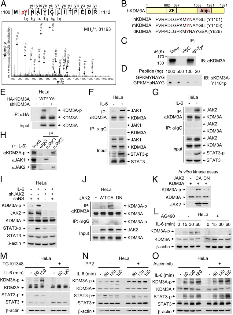
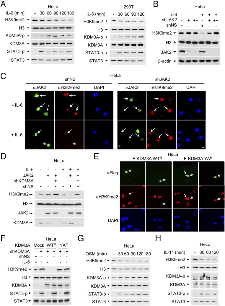
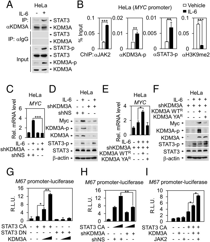
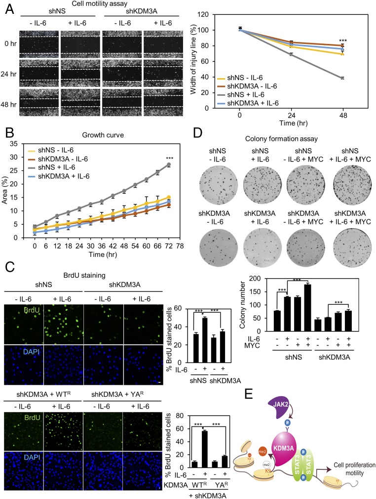
Janus tyrosine kinase 2 (JAK2)-signal transducer and activator of transcription 3 (STAT3) signaling pathway is essential for modulating cellular development, differentiation, and homeostasis. Thus, dysregulation of JAK2-STAT3 signaling pathway is frequently associated with human malignancies. Here, we provide evidence that lysine-specific demethylase 3A (KDM3A) functions as an essential epigenetic enzyme for the activation of JAK2-STAT3 signaling pathway. KDM3A is tyrosine-phosphorylated by JAK2 in the nucleus and functions as a STAT3-dependent transcriptional coactivator. JAK2-KDM3A signaling cascade induced by IL-6 leads to alteration of histone H3K9 methylation as a predominant epigenetic event, thereby providing the functional and mechanistic link between activation of JAK2-STAT3 signaling pathway and its epigenetic control. Together, our findings demonstrate that inhibition of KDM3A phosphorylation could be a potent therapeutic strategy to control oncogenic effect of JAK2-STAT3 signaling pathway.

Keywords: JAK2; KDM3A; STAT3; histone demethylation; phosphorylation.


Conflict of interest statement

The authors declare no conflict of interest.

Figures

Fig. 1.
Fig. 1.
JAK2 phosphorylates KDM3A at tyrosine 1101 residue. (A) Identification by LC-MS/MS of a tyrosine phosphorylation site on KDM3A. (B) Schematic of KDM3A showing zinc finger (ZF) and Jumonji C (Jmjc) domains. Sequences around tyrosine residue (Y, marked by red) are conserved among Drosophila (d), mouse (m), and human (h). (C) Protein extracts from HeLa cells were subjected to pull-down with anti−pan-phospho-tyrosine antibody. Phosphorylation of KDM3A was assessed by immunoblot with anti-KDM3A antibody. (D) Dot blotting of unmodified or phosphorylated KDM3A peptides with anti−phospho-KDM3A (KDM3A-Y1101p) antibody. (E) Endogenous KDM3A was knocked down using shRNA, and an shRNA-resistant form of HA-KDM3A WT (WTR) or HA-KDM3A YA mutant (YAR) was reconstituted in HeLa cells. KDM3A was immunoprecipitated using an anti-HA antibody and analyzed by immunoblot with anti−phospho-KDM3A antibody. (F and G) Co-IP assay of KDM3A with (F) JAK1 or (G) JAK2 was conducted in HeLa cells with or without IL-6 treatment (50 ng/mL) for 2 h. (H) Co-IP assay of JAK1 or JAK2 with phosphorylated KDM3A was conducted in HeLa cells treated with IL-6 for 2 h. Phosphorylation level of KDM3A was assessed by immunoblot analysis using anti−phospho-KDM3A antibody. (I) Endogenous JAK2 was knocked down using shRNA, and phosphorylation level of KDM3A was assessed by immunoblot analysis using anti−phospho-KDM3A antibody in HeLa cells with or without IL-6 treatment for 2 h. (J) WT, constitutively active mutant (CA) or dominant negative mutant (DN) of JAK2 was transfected to HeLa cells. Cell lysates were immunoprecipitated with anti-KDM3A antibody followed by immunoblot analysis with anti−phospho-KDM3A antibody to detect phosphorylated KDM3A. (K) In vitro kinase assay with JAK2 CA or DN mutant using recombinant KDM3A proteins. (LO) HeLa cells, serum-starved for 24 h, were treated with IL-6 (50 ng/mL) for indicated times. Phosphorylation levels of KDM3A were analyzed by immunoblot with anti−phospho-KDM3A antibody in the absence or presence of JAK2 inhibitors (L) AG490 (10 μM) or (M) TG101348 (2.5 μM), (N) an Src family inhibitor PP2 (10 μM), or (O) an Abl inhibitor Asciminib (250 nM).
Fig. 2.
Fig. 2.
KDM3A phosphorylation by JAK2 increases its demethylase activity. (A) The level of H3K9me2 was analyzed in HeLa and HEK293T cells after IL-6 treatment. (B) The level of H3K9me2 was analyzed after knockdown of JAK2 by shRNA in the absence or presence of IL-6. (C) Representative images of cells knocked down by JAK2 shRNA in the absence or presence of IL-6. The cells were stained with anti-H3K9me2 or anti-JAK2 antibodies. Nuclei were counterstained with DAPI. Arrows indicate cells expressing JAK2 and the level of H3K9me2. (Scale bar, 10 μm.) (D) Demethylation of H3K9me2 was induced by JAK2, and knockdown of KDM3A abolished JAK2-dependent demethylation of H3K9me2 in the presence of IL-6. (E) HeLa cells were knocked down by KDM3A shRNA and reconstituted with shRNA-resistant form of KDM3A WT (WTR) or YA (YAR). Transfected cells were fixed and stained for anti-H3K9me2 and anti-Flag antibodies. The cells were counterstained with DAPI to visualize cell nuclei. Arrows indicate cells expressing KDM3A and the level of H3K9me2. (Scale bar, 10 μm.) (F) Demethylation of H3K9me2 induced by IL-6 is dependent on KDM3A. Protein extracts from HeLa cells were knocked down by KDM3A shRNA and reconstituted with KDM3A WTR or YAR. Transfected cells were collected to determine the H3K9me2 levels in the absence or presence of IL-6 by immunoblot analysis. (G and H) The level of H3K9me2 was analyzed after treatment of (G) OSM (20 ng/mL) or (H) IL-11 (5 ng/mL), which share a gp130-mediated signaling molecule in HeLa cells, serum-starved for 24 h.
Fig. 3.
Fig. 3.
KDM3A functions as a transcriptional coactivator of STAT3 in JAK−STAT signaling pathway. (A) Co-IP assay was performed to detect interaction between STAT3 and KDM3A in HeLa cells with or without IL-6 treatment for 2 h. (B) ChIP assays were performed using anti-JAK2, anti−phospho-STAT3, anti−phospho-KDM3A, and anti-H3K9me2 antibodies on the MYC promoters after IL-6 treatment in HeLa cells. **P < 0.01; ***P < 0.001 (Student’s t test). (C) Quantitative RT-PCR analysis of MYC mRNA levels after knockdown of KDM3A by shRNA in HeLa cells following IL-6 treatment. ***P < 0.001 (Student’s t test). (D) Immunoblot analysis of MYC protein levels after knockdown of KDM3A by shRNA following IL-6 treatment in HeLa cells. (E) MYC mRNA levels were measured by quantitative RT-PCR in HeLa cells after rescuing resistant forms of KDM3A WTR or YAR in shRNA-mediated KDM3A knockdown cells following IL-6 treatment. *P < 0.05 (Student’s t test). (F) Immunoblot analysis in HeLa cells after rescuing resistant forms of KDM3A WTR or YAR in shRNA-mediated KDM3A knockdown cells following IL-6 treatment. (GI) STAT-responsive M67 promoter luciferase reporter was transfected into HEK293T cells with indicated plasmids. Luciferase reporter activity was measured at 48 h after transfection and normalized by β-galactosidase activity. Values are expressed as mean ± SD for three independent experiments. *P < 0.05; **P < 0.01 (Student’s t test).
Fig. 4.
Fig. 4.
KDM3A phosphorylation is responsible for increased cell proliferation and motility. (A) (Left) Photomicrographs from the scratch-motility assay of HeLa cells expressing KDM3A shRNA with or without IL-6 treatment. Wound closure was monitored at 24-h intervals for 48 h in HeLa cells. (Right) Cell migration (percent) was quantified by calculating the wound width. ***P < 0.001 (Student’s t test). (B) Proliferation was monitored at 6-h intervals in HeLa cells ectopically expressing either control shRNA or KDM3A shRNA. Proliferation efficiency (percent) was quantified by calculating areas of cell population as shown in the graph. ***P < 0.001 (Student’s t test). (C) Confocal images of cells stained with BrdU. The fraction of increased BrdU-positive (BrdU+) cells after IL-6 treatment decreased following knockdown of KDM3A by shRNA. HeLa cells were knocked down by KDM3A shRNA and reconstituted with an shRNA-resistant form of KDM3A WT (WTR) or YA (YAR). Nuclei were counterstained with DAPI. (Scale bar, 10 μm.) ***P < 0.001 (Student’s t test). (D) (Top) Colony formation assay of HeLa cells transfected with either control shRNA or KDM3A shRNA in combination with MYC with or without IL-6 treatment. Cells were fixed and stained with crystal violet solution. (Bottom) Colony number was quantified as shown in the graph. ***P < 0.001 (Student’s t test). (E) Schematic model depicting JAK2−KDM3A−STAT3 signaling axis. Modulation of H3K9 methylation signature by JAK2-dependent phosphorylation of KDM3A is one of the predominant epigenetic events in transcriptional regulation of STAT3 target genes.

Similar articles

Cited by

References

    1. Bannister AJ, Kouzarides T. Regulation of chromatin by histone modifications. Cell Res. 2011;21:381–395. - PMC - PubMed
    1. Campos EI, Stafford JM, Reinberg D. Epigenetic inheritance: Histone bookmarks across generations. Trends Cell Biol. 2014;24:664–674. - PMC - PubMed
    1. Lee JS, Smith E, Shilatifard A. The language of histone crosstalk. Cell. 2010;142:682–685. - PMC - PubMed
    1. Li B, Carey M, Workman JL. The role of chromatin during transcription. Cell. 2007;128:707–719. - PubMed
    1. Martinez-Jimenez CP, Sandoval J. Epigenetic crosstalk: A molecular language in human metabolic disorders. Front Biosci (Schol Ed) 2015;7:46–57. - PubMed

Publication types

MeSH terms

LinkOut - more resources