. 2018 Oct 30;9(11):1105.
doi: 10.1038/s41419-018-1134-4.

Endoplasmic reticulum resident oxidase ERO1-Lalpha promotes hepatocellular carcinoma metastasis and angiogenesis through the S1PR1/STAT3/VEGF-A pathway

Affiliations

Endoplasmic reticulum resident oxidase ERO1-Lalpha promotes hepatocellular carcinoma metastasis and angiogenesis through the S1PR1/STAT3/VEGF-A pathway

Shikun Yang et al. Cell Death Dis. .

Abstract

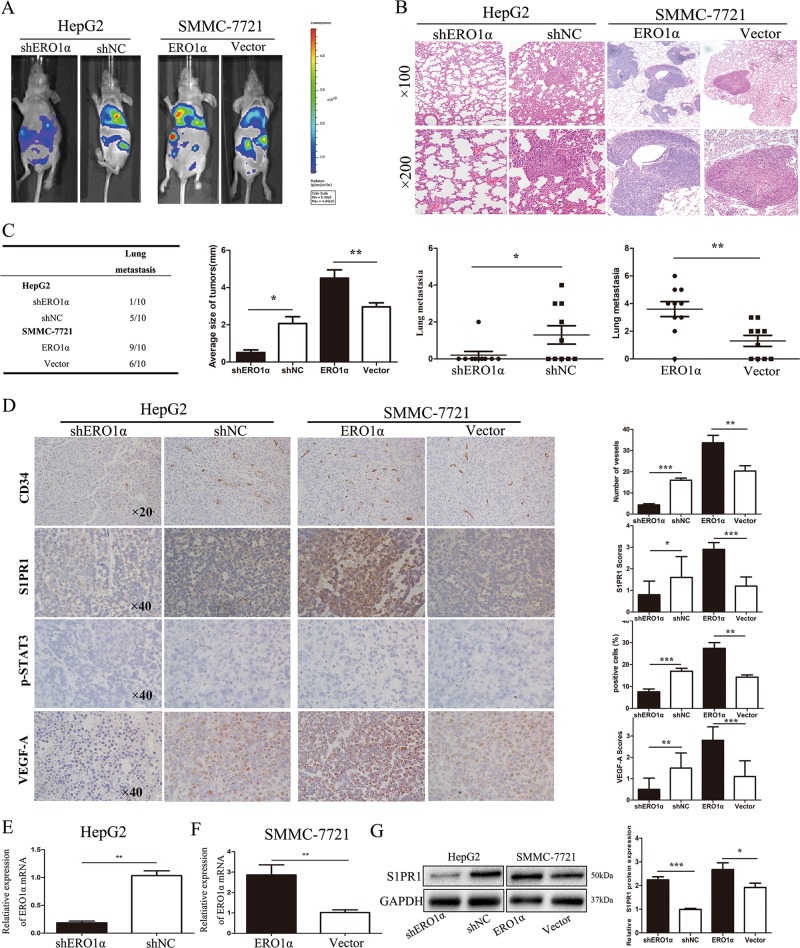
Mounting evidence demonstrates that expression of ERO1α, an endoplasmic reticulum (ER)-resident oxidase, is a poor prognosis factor in a variety of human cancers. However, the clinical relevance of ERO1α and its molecular mechanisms underlying tumor progression have not been determined for hepatocellular carcinoma (HCC). ERO1α expression levels in HCC tissues and cells were detected by quantitative real-time PCR and western blotting. ERO1α shRNAs and overexpression vector were transfected into HCC cells to downregulate or upregulate ERO1α expression. In vitro and in vivo assays were performed to investigate the function of ERO1α in invasion, metastasis, and angiogenesis of HCC. We found high ERO1α expression in HCC tissues and cells that was significantly associated with metastasis and poor clinicopathologic features of vascular invasion, advanced Edmondson Grade, and TNM stage. Loss-of-function and gain-of-function studies showed that ERO1α prompted migration, invasion, epithelial-mesenchymal transition (EMT), and angiogenesis of HCC cells both in vitro and in vivo. Further studies verified a positive correlation between ERO1α and S1PR1, upregulated in metastatic HCC tissues compared with HCC tissues without metastasis. S1PR1 knockdown markedly diminished the effects of ERO1α on HCC cell migration, invasion and vascular endothelial growth factor (VEGF) expression. Most importantly, ERO1α knockdown significantly repressed the death of HCC xenograft mouse models by reducing tumor distant metastasis, and host angiogenesis by suppressing the expression of S1PR1, p-STAT3, and VEGF-A in HCC cells. Our findings suggest that ERO1α is significantly correlated with reduced survival and poor prognosis, and promotes HCC metastasis and angiogenesis by triggering the S1PR1/STAT3/VEGF-A signaling pathway. ERO1α might be a novel candidate in HCC prognosis and therapy.


Conflict of interest statement

The authors declare that they have no conflict of interest.

Figures

Fig. 1
Fig. 1. ERO1α expression is significantly upregulated in HCC tissues samples and cell lines.
a ERO1α mRNA and b protein in HCC tumor tissues compared with adjacent nontumor tissues. c Representative IHC staining of ERO1α in HCC and adjacent tissues. d ERO1α mRNA and e protein in HCC cell lines and normal L02 cells by RT-qPCR and western blotting. *P < 0.05, **P < 0.01, ***P < 0.001
Fig. 2
Fig. 2. Upregulated ERO1α predicts poor prognosis and correlates with metastasis for HCC patients.
a Kaplan–Meier association of higher ERO1α levels with shorter OS and (b) RFS. c ERO1α in HCC with and without metastasis by RT-qPCR. Absence (−) (n = 40) or presence (+) (n = 71) of venous invasion (tumor thrombus in veins of adjacent nontumor tissues or portal vein), lymph node metastasis (n = 2), or both (n = 1). Central horizontal line, mean; error bars, SD. **P < 0.01
Fig. 3
Fig. 3. ERO1α promoted cell migration and invasion of HCC cells in vitro.
a HepG2 and Hep3B cells transfected with shRNA targeting ERO1α were used as shERO1α. Cells transfected with empty lentiviral vectors were negative controls (shNC). ERO1α was detected with RT-qPCR and b western blotting. c Huh-7 and SMMC-7721 cells were transfected for ERO1α overexpression. Cells transfected with empty lentiviral vectors were used as controls. ERO1α was analyzed by RT-qPCR and d western blotting after transfection. e, f, i, j Transwell assays for cell migration and invasion and g, h, k, l wound-healing assays. Cell migration and invasion were quantified as cell numbers. All experiments were performed three times. Data are mean ± SD. *P < 0.05, **P < 0.01, ***P < 0.001
Fig. 4
Fig. 4. ERO1α promoted HCC cell epithelial–mesenchymal transition (EMT) and its ability to promote HUVEC tube formation.
a Western blotting and b immunofluorescent staining for levels of EMT-related proteins with 4′,6-diamidino-2-phenylindole (blue) to identify nuclei. Scale bars = 50 μm. c, e Tube-formation assays with HUVECs in indicated conditioned media. Numbers of branches were calculated using Image Pro Plus 6. d, f Endothelial recruitment assays with HUVECs performed for each group. Cell migration was quantified as cell numbers. All experiments were performed three times. Data are mean ± SD. *P < 0.05, **P < 0.01, ***P < 0.001
Fig. 5
Fig. 5. ERO1α augmented expression of VEGF-A.
a VEGF-A expression analyzed for 114 pairs of HCC tissues from the HCC cohort described in Fig. 2. Central horizontal line, mean; error bars, SD. b Positive correlation between ERO1α and VEGF-A levels in HCC specimens. c VEGF-A protein by western blots. d RT-qPCR for levels of VEGF-A mRNA in tumor cells. e ELISA for VEGF-A concentration in TCM. Data are mean ± SD. *P < 0.05, **P < 0.01, ***P < 0.001
Fig. 6
Fig. 6. S1PR1/STAT3/VEGF-A signaling is critical for ERO1α-mediated promotion of migration, invasion, and angiogenesis.
a S1PR1 expression was analyzed in HCC tissues from the HCC cohort with or without metastasis described in Fig. 2. b Positive correlation between ERO1α and S1PR1 levels in HCC specimens. c Immunofluorescent staining images for ERO1α and S1PR1 in SMMC-7721-ERO1α and SMMC-7721-Vector cells. 4′,6-Diamidino-2-phenylindole (blue) was used to identify nuclei. Scale bars = 50 μm. d Indicated molecules were evaluated by western blots with four HCC cell lines, using GAPDH as the loading control. e RT-qPCR and f western blots for S1PR1 and VEGF-A in indicated cells. hj Rescue experiments for ERO1α-overexpressing cells with S1PR1 silencing. Downregulated S1PR1 counteracted Huh-7 and SMMC-7721 cell migration and invasion that was enhanced by ERO1α overexpression. Cell migration and invasion were quantified as cell numbers. All experiments were performed three in triplicate. Data are mean ± SD. *P < 0.05, **P < 0.01, ***P < 0.001
Fig. 7
Fig. 7. S1PR1/STAT3/VEGF-A pathway contributes to ERO1α-mediated distant metastasis and angiogenesis in an HCC xenograft model.
a Photographs of tumors by an IVIS Imaging System. Representative luciferase signals were captured for each group 7 weeks after injecting indicated cells. b HE-stained lung sections. c, d ERO1α expression in metastatic tumors derived from nude mice measured by RT-qPCR. e S1PR1 protein in metastatic tumors derived from nude mice measured by western blots. f CD34, S1PR1, p-STAT3, and VEGF-A analyzed in tumor tissues by immunohistochemistry. Blood vessels were stained using anti-CD34, and positively stained vessels were counted in five areas per slide to determine maximum number of microvessels; 10 slides per experiment. Results are mean ± SD. *P < 0.05, **P < 0.01, ***P < 0.001

References

    1. Torre L, et al. Global cancer statistics, 2012. CA Cancer J. Clin. 2015;65:87–108. doi: 10.3322/caac.21262. - DOI - PubMed
    1. Siegel R, Miller K, Jemal A. Cancer statistics, 2017. CA Cancer J. Clin. 2017;67:7–30. doi: 10.3322/caac.21387. - DOI - PubMed
    1. Bruix J, Sherman M. Management of hepatocellular carcinoma: an update. Hepatology. 2011;53:1020–1022. doi: 10.1002/hep.24199. - DOI - PMC - PubMed
    1. Yang L, et al. Mesohepatectomy for centrally located large hepatocellular carcinoma: Indications, techniques, and outcomes. Surgery. 2014;156:1177–1187. doi: 10.1016/j.surg.2014.05.012. - DOI - PubMed
    1. Gess B, et al. The cellular oxygen tension regulates expression of the endoplasmic oxidoreductase ERO1-Lalpha. Eur. J. Biochem. 2003;270:2228–2235. doi: 10.1046/j.1432-1033.2003.03590.x. - DOI - PubMed

Publication types

MeSH terms